Tag Archives: INTERNATIONAL ASSOCIATION OF HEALTHCARE PROFESSIONALS

World watches as grandfather from Malawi has his sight restored live online

Ophthalmology

A blind man’s sight-restoring operation was broadcast live around the world at 1.30pm (BST) October 8, 2014 from a hospital in Malawi.

The six minute cataract operation will mean 69-year-old Winesi March could see his baby grandson for the first time when his bandages are removed tomorrow on World Sight Day.

The live online broadcast was hosted by YouTuber Doug Armstrong who fielded questions from the global audience via a Google Hangout. Dr Gerald Msukwa, one of only a few ophthalmologists in Malawi, talked through the simple procedure whichrestores the sight of more than 20 million people around the world every year and is the most commonly performed surgery on the NHS.

Dr Msukwa said:

“I’m a doctor, not a movie star so there is some tension with the world watching but it’s nothing when I know that tomorrow my patient’s life will be utterly changed.

“Yesterday Winesi could not farm his land, see his family or walk to the market without the constant support of his dedicated wife. Tomorrow he tells me he will dance across the river by his home to work. The operation only costs GBP30 but will help him feed his family for years to come.”

The second live broadcast (October 9, 2014) will see the global audience join the team in Malawi for the life-changing moment when Winesi’s bandages are removed and he sees his 18-month-old grandson Luka for the first time.

In the UK more than 50 per cent of adults over the age of 65 have cataract, a condition that causes sight to become blurred and gradually lost. But the majority of the 20 million people blind from cataracts are living in the poorest parts of the world, often unable to access the straightforward surgery.

Winesi’s surgery is the first ‘miracle’ of Sightsavers’ biggest-ever fundraising appeal – A Million Miracles. The charity is aiming to raise GBP30 million to provide one million sight-restoring surgeries for people living in developing countries. All donations made by the UK public will be matched pound for pound by the UK government for the first three months of the appeal.

To watch the online surgery again go to millionmiracles.org


Retinal tumors in young children caused by a single, genetic change

Ophthalmology_Oncology_Pediatrics

Retinoblastoma is a childhood retinal tumor usually affecting children one to two years of age. Although rare, it is the most common malignant tumor of the eye in children. Left untreated, retinoblastoma can be fatal or result in blindness. It has also played a special role in understanding cancer, because retinoblastomas have been found to develop in response to the mutation of a single gene – the RB1 gene – demonstrating that some cells are only a step away from developing into a life-threatening malignancy.

David E. Cobrinik, MD, PhD, of The Vision Center at Children’s Hospital Los Angeles (CHLA), together with colleagues at Memorial Sloan-Kettering Cancer Center, has answered the long-standing question of why mutations to the RB1 gene primarily cause tumors of the retina and not of other cell types. His study – which could reveal new cellular signaling pathways relevant to retinal development, cancer development, and ultimately, the development of novel therapies – is published in this week’s early on line issue of the journal Nature.

“These findings significantly advance our understanding of cancer, not only because they solve the RB riddle, but also because they more generally imply that cancers can develop through the collaboration between a cancer-causing mutation – in this case, inactivation of the RB1 gene – and cell type-specific circuitry,” said Cobrinik, who also an investigator with The Saban Research Institute of CHLA and associate professor of Ophthalmology at USC Eye Institute, Keck School of Medicine at the University of Southern California.

The RB1 gene encodes a tumor suppressor protein, referred to as Rb, which prevents excessive cell growth by inhibiting cell cycle progression until a cell is ready to divide. If both alleles of the RB1 gene are mutated early in life, the Rb protein is inactivated, resulting in development of retinoblastoma cancers. (While the Rb protein regulates proliferation in many cell types, only cells in the retina routinely form cancers when the function of the RB1 gene is lost.)

Cobrinik and colleagues discovered that retinoblastomas originate in cone photoreceptor precursors, and their study explains why retinoblastomas originate in these precursor cells. Cone cells, or cones, are one of the two types of photoreceptor cells in the retina, and are responsible for color vision. A cone precursor is an immature cone cell which is not yet fully differentiated.

The study indicates that cone precursors prominently express key, cancer-related proteins that enable proliferation and suppress apoptosis, or programmed cell death. Meanwhile, the role of the Rb protein is to hold back such proliferation – which means that the loss of Rb alone is sufficient to allow unchecked cell proliferation, causing retinoblastomas to form.

“We showed that the cone precursors’ normal developmental program collaborates with RB1 mutations to deregulate cell growth,” Cobrinik explained. “In other words, loss of the RB1 gene results in abnormal proliferation because the cone precursor cells lack a self-monitoring ‘surveillance system’ – which would normally cause aberrantly proliferating cells to undergo apoptosis. Instead, cells are able to divide uncontrollably and eventually become cancerous.”

http://www.medicalnewstoday.com/releases/283008.php


Brain mechanism underlying the recognition of hand gestures develops even when blind

Ophthalmology

Does a distinctive mechanism work in the brain of congenitally blind individuals when understanding and learning others’ gestures? Or does the same mechanism as with sighted individuals work? Japanese researchers figured out that activated brain regions of congenitally blind individuals and activated brain regions of sighted individuals share common regions when recognizing human hand gestures. They indicated that a region of the neural network that recognizes others’ hand gestures is formed in the same way even without visual information. The findings are discussed in The Journal of Neuroscience (July 23, 2014 electronic edition).

Our brain mechanism perceives human bodies from inanimate objects and shows a particular response. A part of a region of the “visual cortex” that processes visual information supports this mechanism. Since visual information is largely used in perception, this is reasonable, however, for perception using haptic information and also for the recognition of one’s own gestures, it has been recently learned that the same brain region is activated. It came to be considered that there is a mechanism that is formed regardless of the sensory modalities and recognizes human bodies.

Blind and sighted individuals participated in the study of the research group of Assistant Professor Ryo Kitada of the National Institute for Physiological Sciences, National Institutes of Natural Sciences. With their eyes closed, they were instructed to touch plastic casts of hands, teapots, and toy cars and identify the shape. As it turned out, sighted individuals and blind individuals could make an identification with the same accuracy. Through measuring the activated brain region using functional magnetic resonance imaging (fMRI), for plastic casts of hands and not for teapots or toy cars, the research group was able to pinpoint a common activated brain region regardless of visual experience. On another front, it also revealed a region showing signs of activity that is dependent on the duration of the visual experience and it was also learned that this region functions as a supplement when recognizing hand gestures.

As Assistant Professor Ryo Kitada notes, “Many individuals are active in many parts of the society even with the loss of their sight as a child. Developmental psychology has been

advancing its doctrine based on sighted individuals. I wish this finding will help us grasp how blind individuals understand and learn about others and be seen as an important step in supporting the development of social skills for blind individuals.”

http://www.medicalnewstoday.com/releases/282166.php


DNA test for congenital cataracts leads to faster, more accurate diagnoses of rare diseases linked to childhood blindness

Ophthalmology_Pediatrics

Researchers in the United Kingdom have demonstrated that advanced DNA testing for congenital cataracts can quickly and accurately diagnose a number of rare diseases marked by childhood blindness, according to a study published online in Ophthalmology, the journal of the American Academy of Ophthalmology. Using a single test, doctors were able to tailor care specifically to a child’s condition based on their mutations reducing the time and money spent on diagnosis and enabling earlier treatment and genetic counseling.

Each year, between 20,000 and 40,000 children worldwide are born with congenital cataracts, a disease that clouds the lens of the eye and often requires surgery and treatment to prevent blindness.[1] The disease can arise following a maternal infection or be inherited as an isolated abnormality. Congenital cataracts can also appear as a symptom of more than 100 rare diseases, making mutations in the 115 genes associated with congenital cataracts useful as diagnostic markers for the illnesses.

Diagnosing these rare diseases previously proved a lengthy, costly and inconclusive process involving numerous clinical assessments and taking a detailed family history. DNA testing, one gene at a time, would have taken years to complete. Employing new DNA sequencing technology, called targeted next-generation sequencing, researchers at the University of Manchester sped up diagnosis to a matter of weeks by testing for mutations in all 115 known congenital cataracts genes at one time.

In 75 percent of the 36 cases tested, the DNA test determined the exact genetic cause of congenital cataracts. In one case, the DNA test helped diagnose a patient with Warburg Micro syndrome, an extremely rare disease that is marked by an abnormally small head and the development of severe epilepsy, among other medical issues. Having a clear diagnosis allowed for genetic counseling and appropriate care to be delivered quicker than previously possible without the test.

“There are many diseases that involve congenital cataracts but finding the exact reason was always difficult,” said Graeme Black, DPhil., professor of genetics and ophthalmology at the University of Manchester and strategic director of the Manchester Centre for Genomic Medicine. “Even with a family history, diagnosing these rare diseases was always a bit of a shot in the dark.”

In the course of their work, done in collaboration with Manchester Royal Eye Hospital, researchers also found previously undescribed mutations linked to cataract formation. “There is hope that our work may one day provide more insight into the development and treatment of age-related cataracts, a leading cause of blindness worldwide,” said Rachel Gillespie, MSc, lead author of the study who designed and developed the test.

The test was made available to U.K. patients through the country’s National Health Service in December 2013. Infants and children who have congenital cataracts can be tested as well as prospective parents with a history of the condition who wish to evaluate the risk to their child. Results generally take about two months. While only available in the U.K., the congenital cataract DNA test can be requested by registered medical facilities through international referral.

As with all genetic testing, the American Academy of Ophthalmology encourages clinicians and patients to consider the benefits as well as the risks. Ophthalmologists who order genetic tests either should provide genetic counseling to their patients themselves, if qualified to do so, or should ensure that counseling is provided by a trained individual, such as a board-certified medical geneticist or genetic counselor. For more information, please see the Academy’s recommendations on genetic testing for inherited eye diseases.


http://www.medicalnewstoday.com/releases/281442.php


Slippery material for lubricating joints inspired by nature

Ophthalmology_Orthopedic Surgery

For some time, scientists have been aware that synovial fluid in joints contains a natural substance that helps keep them well lubricated. Now, by mimicking its properties, engineers at Johns Hopkins University in Baltimore, MD, hope to develop a new material that delivers long-lasting lubrication in artificial joints, and other specific spots in the body where surfaces move against each other.

The team writes about the new material, and the potential it offers, in the journal Nature Materials. As well as helping to ease joint pain, other areas of possible use include making contact lenses more comfortable.

The molecule they are investigating is called hyaluronic acid (HA), which exists in several forms in the body where lubrication is needed.

One form of HA reduces inflammation and protects cells from metabolic damage. In the body, HA is bound to the surfaces it protects by a protein. Research shows that in damaged, diseased and aging knees, hips, shoulders and elbows, this protein is no longer able to hold onto HA.

Viscosupplementation is a popular treatment for painful joints and consists of injecting HA into the painful joint. However, if the cause of the pain is the lack of the protein that helps bind HA to the affected surface, then it does not offer long-lasting benefit. The injected HA is soon washed away by the body’s natural cleaning processes.

‘Chemical handle’ holds HA in place

Thus, led by Jennifer H. Elisseeff, a professor at the Wilmer Eye Institute at Johns Hopkins, the team sought to find a way to keep HA in place. They found their answer in molecules known as HA-binding peptides (HABpeps).

The scientists used HABpep as a “chemical handle” to attach HA onto natural and artificial surfaces with the help of another synthetic molecule, polyethylene glycol.

In the lab, they tested the new material in cultured tissue and joint and eye surface tissue in live animals. They found the bound HA did not wash away easily, and it reduced friction as well as when the tissues were immersed in a bath of HA:

“Tissue surfaces treated with the HA-binding system exhibited higher lubricity values, and in vivo were able to retain HA in the articular joint and to bind ocular tissue surfaces,” they note.

HABpep holds HA in place 12 times longer

In another set of experiments, they also tested an HABpep designed to attach to cartilage. They injected rats’ knees first with some HABpep, then with some HA, and found the HA stayed in place 12 times longer than it did in rats that had received only HA injections without HABpep.

The team suggests this shows HABpeps may be a useful addition to viscosupplementation by helping the HA to stay in place longer. They also conclude:

“Biomaterials-mediated strategies that locally bind and concentrate HA could provide physical and biological benefits when used to treat tissue-lubricating dysfunction and to coat medical devices.”

Although it will be some time before such a material is ready for use in humans, the team highlights that theirs is yet another example of where nature has inspired the solution to a medical problem.

Prof. Elisseeff, who is also of the Johns Hopkins University departments of Biomedical Engineering and of Materials Science and Engineering, says:

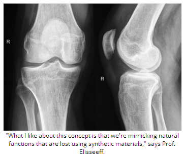
“What I like about this concept is that we’re mimicking natural functions that are lost using synthetic materials.”

Meanwhile in November 2013, Medical News Today learned how another team of researchers – also inspired by nature – is refining sea coral for use in bone grafts.

Written by Catharine Paddock PhD

http://www.medicalnewstoday.com/articles/281212.php


Age-related macular degeneration occurs much earlier than previously assumed

Ophthalmology

It is widely accepted that age-related macular degeneration (AMD) is the most common cause of visual impairment and blindness in industrialized countries. However, it is questionable whether it can continue to be defined as a disease in people in their 50s and beyond. Investigations to determine the incidence of age-related macular degeneration undertaken as part of the Gutenberg Health Study of the University Medical Center of Johannes Gutenberg University Mainz (JGU) have shown that even persons under the age of 50 years may be affected by an early form of the eye disease. Just under 4 percent of the 35 to 44-year-old subjects in the population-based study were found to be suffering from AMD.

In order to identify the age- and gender-specific incidence of AMD, the research team of the Department of Ophthalmology at the Mainz University Medical Center led by Dr. Christina Korb, PD Dr. Alireza Mirshahi, and Professor Norbert Pfeiffer assessed the status of the ocular fundus of 4,340 participants in the Gutenberg Health Study. Evaluated were vascular structure, the head of the optic nerve, and the macula of the eye, which is the point of sharpest vision. The results in general documented that the incidence of AMD increases with age. However, the researchers also discovered to their surprise that even persons under the age of 50 years can already be affected by early stage AMD. In the age group of 35- to 44-year-olds, 3.8 percent of the subjects in the Gutenberg Health Study were found to be suffering from the disease. The findings of the Mainz researchers thus contradict the current assumption that age-related macular degeneration only occurs in the section of the population that is over 50 years old.

With the help of their findings, the researchers were also able to gain insights into how frequently the various forms of age-related macular degeneration occur. On average, about 12 percent of the examined 35- to 74-year-olds had early stage AMD, but only 0.2 percent of the study participants exhibited symptoms of late stage AMD, which is often associated with severe visual impairment. “Our research shows that age-related macular degeneration can already occur much earlier than previously thought. This means there may also be possible consequences with regard to the screening examinations for these diseases,” concluded Dr. Christina Korb.

Age-related macular degeneration leads to loss of visual acuity. The cause is damage to the cells in the region of the central retina also known as the “yellow spot.” There is still insufficient information on the annual number of individuals who develop AMD and the Mainz-based researchers hope to be able to remedy this with the help of their next project. As the Gutenberg cohort was subjected to a follow-up examination five years after inclusion in the study, the research group has now access to more relevant and reliable data. “The prospective design of the study, in combination with the availability of interdisciplinary research data, should make it possible for us to identify risk factors for the development of late forms of AMD in our cohort. We are looking forward with some excitement to the results,” explained the team. The objective is to reveal, for the first time, the incidence of AMD across the whole population of Germany.

The Gutenberg Health Study (GHS) is an interdisciplinary, population-based, prospective, monocenter cohort study, which has been conducted at the Mainz University Medical Center since 2007. Cardiovascular diseases,cancer, eye diseases, metabolic disorders as well as immune system and mental disorders are being investigated as part of the study. The goal of the study is to improve the individual risk prediction for these diseases. To this end, lifestyle, psychosocial factors, environment, clinical laboratory parameters, and the severity of any subclinical disorder are being taken into consideration. A comprehensive biorepository is being developed so that molecular biological investigations can be conducted. During the baseline visit, 15,010 participants aged 35 to 74 years were invited to participate in a 5-hour examination program at the study center. This was followed by a computer-assisted telephone interview (CATI) using a standardized questionnaire and the assessment of diseases and health problems after 2.5 years. All endpoints will be subjected to extensive validation. In April 2012, a detailed follow-up examination of participants similar to the baseline examination was conducted at the center five years after their inclusion in the study. The aim is to continue to monitor the cohort and conduct further tests.

http://www.medicalnewstoday.com/releases/279917.php

Picture courtesy of lighthouse.org


Computer Use and Eye Strain

Staring at your computer screen, smartphone, video game or other digital devices for long periods won’t cause permanent eye damage, but your eyes may feel dry and tired. Some people also experience headaches or motion sickness when viewing 3-D, which may indicate that the viewer has a problem with focusing or depth perception.

What causes computer-use eyestrain?

Woman looking at laptop
  • Normally, humans blink about 18 times a minute, but studies show we blink half that often while using computers and other digital screen devices, whether for work or play.
  • Extended reading, writing or other intensive “near work” can also cause eyestrain.

What to do:

  • Sit about 25 inches from the computer screen and position the screen so your eye gaze is slightly downward.
  • Reduce glare from the screen by lighting the area properly; use a screen filter if needed.
  • Post a note that says “Blink!” on the computer as a reminder.
  • Every 20 minutes, shift your eyes to look at an object at least 20 feet away, for at least 20 seconds: the “20-20-20” rule.
  • Use artificial tears to refresh your eyes when they feel dry.
  • Take regular breaks from computer work, and try to get enough sleep at night.

Computer-use eyestrain can be made worse by:

Sleep deprivation. When you get less sleep than you need, your eyes may become irritated. During sleep our eyes rest for an extended period and are replenished by nutrients. Ongoing eye irritation can lead to swelling and infection, especially if you wear contact lenses.

  • If you have to be at your computer for a marathon work session, take regular rest breaks or “power naps,” if possible.
  • Apply a washcloth soaked in warm water to tired, dry eyes (with eyes closed).
  • Use tired or sore eyes as a signal that it’s time to stop working and get some rest or sleep.

Incorrect contact lens use. If you wear contact lenses, it’s important that you use and care for them properly — especially if you use a computer and other digital-screen devices often. This helps avoid eye irritation, swelling, infection and vision problems.

  • Give your eyes a break: wear your glasses!
  • Don’t sleep in your contact lenses, even if they are labeled “extended wear.”
  • Always use good cleaning practices.

There are some important things to keep in mind when cleaning your contact lenses. You should:

  • Avoid touching the lenses with water; use fresh solution every time for cleaning and storing.
  • Rub your contacts when you clean them, even if you use a no-rub solution.
  • Clean your storage case regularly (with fresh solution, not water) and replace it every 2 to 3 months.

Stop wearing your contact lenses and see an ophthalmologist (Eye M.D.) right away if you develop any of these problems: Eyes that are red, blurry, watery, sensitive to light, or sore; eye swelling or discharge.

http://www.geteyesmart.org/eyesmart/living/computer-usage.cfm

Myths and Facts About Ophthalmology

Myth

Reading in dim light is harmful to your eyes.

Fact

Although reading in dim light can make your eyes feel tired, it is not harmful.

Myth

It is not harmful to watch a welder or look at the sun if you squint, or look through narrowed eyelids.

Fact

Even if you squint, ultra-violet light still gets to your eyes, damaging the cornea, lens, and retina. Never watch welding without wearing the proper protection. Never look directly at an eclipse.

Myth

Using a computer, or video display terminal (VDT), is harmful to the eyes.

Fact

Although using a VDT is associated with eyestrain or fatigue, it is not harmful to the eyes.

Myth

If you use your eyes too much, you wear them out.

Fact

You can use your eyes as much as you wish – they do not wear out.

Myth

Wearing poorly-fit glasses damages your eyes.

Fact

Although a good fit is required for good vision with glasses, a poor fit does not damage your eyes.

Myth

Wearing poorly fit contacts does not harm your eyes.

Fact

Poorly fit contact lenses can be harmful to your cornea (the window at the front of your eye). Make certain your eyes are checked regularly by an eye doctor at our ophthalmology office if you wear contact lenses.

Myth

You do not need to have your eyes checked until you are in your 40s or 50s.

Fact

There are several asymptomatic, yet treatable, eye diseases (most notably glaucoma) that can begin prior to your 40s.

Myth

Safety goggles are more trouble than they’re worth.

Fact

Safety goggles prevent many potentially blinding injuries every year. Keep goggles handy and use them.

Myth

It’s okay to swim while wearing soft contact lenses.

Fact

Potentially blinding eye infections can result from swimming or using a hot tub while wearing contact lenses.

Myth

Children outgrow crossed eyes.

Fact

Children do not outgrow truly crossed eyes. A child whose eyes are misaligned has strabismus and can develop poor vision in one eye (a condition known as amblyopia) because the brain turns off the misaligned or “lazy” eye. The sooner crossed or misaligned eyes are treated, the less likely the child will have permanently impaired vision.

Myth

A cataract must be ripe before it can be removed.

Fact

With modern cataract surgery, a cataract does not have to ripen before it is removed. When a cataract keeps you from doing the things you like or need to do, consider having it removed.

Myth

Cataracts can be removed with lasers.

Fact

A cataract cannot be removed with a laser. The cloudy lens must be removed through a surgical incision. However, after cataract surgery, a membrane within the eye may become cloudy. This membrane can be opened with laser eye surgery.

Myth

Eyes can be transplanted.

Fact

The eye cannot be transplanted. It is connected to the brain by the optic nerve, which cannot be reconnected once it has been severed. A person can, however, undergo cornea transplant surgery to replace the cornea, the clear part of the eye. Surgeons often use plastic intraocular lens implants (IOLs) such as crystalens®, ReSTOR®, ReZoom™ or TECNIS® to replace natural lenses during cataract surgery.

http://www.batravision.com/html/eye-care.html

Renee C. Bovelle, MD, FAAO, Represents Maryland with Entry into Renowned Publication The Leading Physicians of the World

Image

The Leading Physicians of the World and the International Association of Healthcare Professionals are proud to announce that Renee C. Bovelle, MD, FAAO, is being honored for her exceptional successes in the ophthalmology community.  Over 15 years practicing, Dr. Bovelle has already demonstrated the passion, dedication, and enthusiasm for patient care necessary to be considered a Top Physician in her field.

Dr. Bovelle currently maintains her own practice, Envision Eye and Laser Center, located in Glenn Dale, Maryland. Within her practice, Dr. Bovelle has performed thousands of LASIK, laser, ophthalmic, cataract and BOTOX procedures; with specific specialization in cornea and refractive surgery. Certified by the American Board of Ophthalmology, she offers both non-surgical and surgical treatments to enhance and rejuvenate her patients; as well as individualized treatment plans to help her patients make informed decisions about their medical, surgical, and aesthetic treatments. Her passion for the field goes beyond her practice, Dr. Bovelle’s work includes clinical research on modern devices used in modern refractive surgery; having her findings published in medical textbooks and ophthalmology journals in the United States and internationally. She also lectures across the globe. Dr. Bovelle also donates her time to the underprivileged both locally and internationally by participating in health fairs and medical mission trips including in Prince George’s County, Washington DC, Jamaica, Africa and India. Also being an advocate for continued education, Dr. Bovelle attends additional training courses in the disciplines of oculo-plastics and aesthetics; as well as, served as an Assistant Professor of Ophthalmology at Louisiana State University, and has worked at the National Institutes of Health.

Dr. Bovelle’s career began in 1992, after she received her medical degree from the University of California in Los Angeles. She completed her internships at a Johns Hopkins-affiliated hospital and at the MedStar Franklin Square Medical Center.  Her residency in ophthalmology was completed at the esteemed Yale University, where she served as chief resident. Upon completion, Dr. Bovelle attended the Louisiana State University Eye Center for a Cornea and Refractive Surgery fellowship under the direction of Dr. Herbert Kaufman, one of the world’s leading corneal and refractive surgeons.

To find out more about Dr. Bovelle, please visit www.renee-bovelle.findatopdoc.com andwww.envisioneyeandlaser.com.  Be sure to look out for her upcoming publication in The Leading Physicians of the World.