Tag Archives: eye surgeon

A pinch of baking soda for better vision?

OphthalmologyBicarbonate (baking soda) makes sparkling water sparkle, causes bread to rise, absorbs odors and can be used for cleaning all sorts of stuff, including your teeth. In the body, it plays essential roles in buffering pH, aiding in digestion and neutralizing lactic acid produced during physical exertion. Much of the bicarbonate in our bodies comes from carbon dioxide, which is produced as a waste product in all cells, although some is ingested with carbonated beverages and certain types of foods.Now a new study from the Makino Laboratory at Massachusetts Eye and Ear/Harvard Medical School and colleagues at Salus University, describes how bicarbonate also alters how we see by modifying the visual signal generated by rod and cone photoreceptors that detect light. This study is described online in the Journal of Biological Chemistry.Within rods and cones, a small soluble molecule, cGMP, links photon absorption to the electrical activity of the photoreceptor. In the light, cGMP is destroyed and ion channels are closed. Positively charged sodium ions cease to enter the rod or cone and the membrane potential becomes more negative or hyperpolarized. Bicarbonate directly stimulates an enzyme called guanylate cyclase that synthesizes cGMP.

Read the rest of the article at http://www.medicalnewstoday.com/releases/291082.php.

Mystery of the reverse-wired eyeball solved

OphthalmologyFrom a practical standpoint, the wiring of the human eye – a product of our evolutionary baggage – doesn’t make a lot of sense. In vertebrates, photoreceptors are located behind the neurons in the back of the eye – resulting in light scattering by the nervous fibers and blurring of our vision. Recently, researchers at the Technion – Israel Institute of Technology have confirmed the biological purpose for this seemingly counterintuitive setup.”The retina is not just the simple detector and neural image processor, as believed until today,” said Erez Ribak, a professor at the Technion – Israel Institute of Technology. “Its optical structure is optimized for our vision purposes.” Ribak and his co-authors will describe their work during the 2015 American Physical Society March Meeting, on Thursday, March 5 in San Antonio, Texas.


Read the rest of the article at http://www.medicalnewstoday.com/releases/290150.php.


Easy on the eyes: How eyelash length keeps your eyes healthy

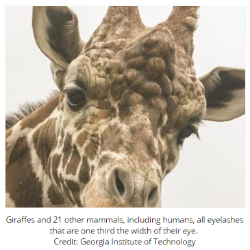
OphthalmologyStudy finds that animals and humans have similar lash length.It started with a trip to the basement of the American Museum of Natural History in New York to inspect preserved animal hides. Later, Georgia Institute of Technology researchers built a wind tunnel about 2 feet tall, complete with a makeshift eye. By putting both steps together, the team discovered that 22 species of mammals – from humans, to hedgehogs, to giraffes ¬- are the same: their eyelash length is one-third the width of their eye. Anything shorter or longer, including the fake eyelashes that are popular in Hollywood and make-up aisles, increases airflow around the eye and leads to more dust hitting the surface.”Eyelashes form a barrier to control airflow and the rate of evaporation on the surface of the cornea,” said Guillermo Amador, a Georgia Tech Ph.D. candidate in the George W. Woodruff School of Mechanical Engineering who authored the study. “When eyelashes are shorter than the one-third ratio, they have only a slight effect on the flow. Their effect is more pronounced as they lengthen up until one-third. After that, they start funneling air and dust particles into the eye.”

Read the rest of the article at http://www.medicalnewstoday.com/releases/289941.php.

Attention! How eyes reveal the brain’s focus

OphthalmologyThe eyes are windows into the attention span at least Whether you’re taking a test or walking your dog across a busy street, your ability to tune out irrelevant sights and sounds in the environment — or your openness to detecting potential dangers — is crucial for success and survival.Duke University researchers have looked into monkeys’ eyes for insight into how the brain processes distractions and they’ve found that changes to pupil size in response to distractors might predict how well the brain focuses on a goal.The results, appearing in the journal Neuron, may inform our understanding of attention deficit hyperactivity disorder or other disorders in which mechanisms for maintaining attention go awry. The results could also inspire new ways to improve performance in school or on the job.In recent years, researchers have gained a greater appreciation of the importance of eye movements and pupil size for focusing the mind, and how the brain might regulate these behaviors.

Read the rest of the article at http://www.medicalnewstoday.com/releases/289028.php.

Scientists map brains of the blind to solve mysteries of human brain specialization

Ophthalmology_Neurology

New research suggests unexpected brain connectivity can lead to fast brain specialization, allowing humans to adapt to the rapid technological and cultural innovation of our generation

Studying the brain activity of blind people, scientists at the Hebrew University of Jerusalem are challenging the standard view of how the human brain specializes to perform different kinds of tasks, and shedding new light on how our brains can adapt to the rapid cultural and technological changes of the 21st Century.

Research Highlights:

  • Understanding the brain activity of the blind can help solve one of the oddest phenomena in the human brain: how can tasks such as reading and recognizing numerical symbols have their own brain regions if these concepts were only developed several thousand years ago (which is negligible on an evolutionary timescale)? What was the job of these regions before their invention?
  • New research published in Nature Communicationsdemonstrates that vision is not a prerequisite for “visual” cortical regions to develop these preferences.
  • This stands in contrast to the current main theory explaining this specialization, which suggests these regions were adapted from other visual tasks such as the angles of lines and their intersections.
  • These results show that the required condition is not sensory-based (vision) but rather connectivity- and processing-based. For example, blind people reading Braille using their fingers will still use the “visual” areas.
  • This research uses shows unique connectivity patterns between the visual-number-form-area (VNFA) to quantity-processing areas in the right hemisphere, and between the visual-word-form-area (VWFA) to language-processing areas in the left hemisphere.
  • This type of mechanism can help explain how our brain adapts quickly to the changes of our era of constant cultural and technological innovations.

The accepted view in previous decades was that the brain is divided into distinct regions mainly by the sensory input that activates them, such as the visual cortex for sight and the auditory cortex for sound. Within these large regions, sub-regions have been defined which are specialized for specific tasks such as the “visual word form area,” a functional brain region believed to identify words and letters from shape images even before they are associated with sounds or meanings. Similarly there is another area that specializes in number symbols.

Now, a series of studies at the Hebrew University’s Amedi Lab for Brain and Multisensory Research challenges this view using unique tools known as Sensory Substitution Devices (SSDs). Sensory Substitution Devices take information from one sense and present it in another, for example enabling blind people to “see” by using other senses such as touching or hearing. By using a smartphone or webcam to translate a visual image into a distinct soundscape, SSDs enable blind users to create a mental image of objects, such as their physical dimensions and color. With intense training, blind users can even “read” letters by identifying their distinct soundscape.

“These devices can help the blind in their everyday life,” explains Prof. Amir Amedi, “but they also open unique research opportunities by letting us see what happens in brain regions normally associated with one sense, when the relevant information comes from another.”

Amedi’s team was interested in whether blind subjects using sensory substitution would, like sighted people, use the visual-word-form-area sub-region of the brain to identify shape images, or whether this area is specialized exclusively to visual reading with the eyes.

In a new paper published in Nature Communications as “A number-form area in the blind,” Sami Abboud and colleagues in the Amedi Lab show that these same “visual” brain regions are used by blind subjects who are actually “seeing” through sound. According to lead researcher Sami Abboud, “These regions are preserved and functional even among the congenitally blind who have never experienced vision.”

The researchers used functional MRI imaging (fMRI) to study the brains of blind subjects in real-time while they used an SSD to identify objects by their sound. They found that when it comes to recognizing letters, body postures and more, specialized brain areas are activated by the task at hand, rather than by the sense (vision or hearing) being used.

The Amedi team examined a recently-identified area in the brain’s right hemisphere known as the ‘Visual Number Form Area.’ The very existence of such an area, as distinct from the visual-word-form-area, is surprising since symbols such as ‘O’ can be used either as the letter O or as the number Zero, despite being visually identical.

Previous attempts to explain why both the word and number areas exist, such as the ‘Neural recycling theory’ by Dehaene and Cohen (2007), suggest that in the far distant past these areas were specialized for other visual tasks such as recognizing small lines, their angles and intersections, and thus were best suited for them. However, this new work shows that congenitally blind users using the sensory substitution devices still have these exact same areas, suggesting that vision is not the key to their development.

“Beyond the implications for neuroscience theory, these results also offer us hope for visual rehabilitation,” says Amedi. “They suggest that by using the right technology, even non-invasively, we can re-awaken the visually deprived brain to process tasks considered visual, even after many years of blindness.”

But if the specific sensory input channel is not the key to developing these brain regions, why do these functions develop in their specific anatomical locations? The new research points to unique connectivity patterns between the visual-word-form-area and language-processing areas, and between the visual-number-form-area and quantity-processing areas.

Amedi suggests, “This means that the main criteria for a reading area to develop is not the letters’ visual symbols, but rather the area’s connectivity to the brain’s language-processing centers. Similarly a number area will develop in a region which already has connections to quantity-processing regions.”

“If we take this one step further,” adds Amedi, “this connectivity-based mechanism might explain how brain areas could have developed so quickly on an evolutionary timescale. We’ve only been reading and writing for several thousand years, but the connectivity between relevant areas allowed us to create unique new centers for these specialized tasks. This same ‘cultural recycling’ of brain circuits could also be true for how we will adapt to new technological and cultural innovations in the current era of rapid innovation, even approaching the potential of the Singularity.”

Adapted by MNT from original media release

http://www.medicalnewstoday.com/releases/288490.php


Echolocation: a ‘sixth sense’ for blind people

Ophthalmology

Could echolocation – a skill commonly linked with bats – be used as a “sixth sense” for blind people? Researchers have found that blind people using echolocation are just as susceptible to visual illusions as sighted people.


Echolocation is a process whereby sound waves are used to determine the location and size of objects within a particular area. Returning sound waves rebounding from objects provide positional information to those that are unable to see them.

In the new study, published in Psychological Science, researchers from the Brain and Mind Institute at Western University, Canada, demonstrate that echolocation works in partnership with other senses to provide information to people with visual impairment.

“Some blind people use echolocation to assess their environment and find their way around,” says study author Dr. Gavin Buckingham. “They will either snap their fingers or click their tongue to bounce sound waves off objects, a skill often associated with bats, which use echolocation when flying.”

Researchers found that blind participants in the study were susceptible to the size-weight illusion – also known as the Charpentier illusion – when using echolocating techniques such as snapping their fingers or clicking their tongues while making a judgement about the weights of different sized cubes.

The size-weight illusion is most frequently explained with the example of two boxes – a small one containing 1 kg of iron and a large one containing 1 kg of feathers. The size-weight illusion causes people to perceive the smaller box as heavier, despite them both having the same mass.

“Ironically, the proof for the vision-like qualities of echolocation came from blind echolocators wrongly judging how heavy objects of different sizes felt,” says Melvyn Goodale, director of the Brain and Mind Institute.

Succumbing to the size-weight illusion

For the study, participants were asked to judge the weight of three cubes. The cubes all shared the same weight but were differently sized and could only be lifted by the participants pulling a string attached to the top of each box.

Participants were split into three different groups: blind people using echolocation, blind people not using echolocation, and a group of participants without any visual impairment.

People from the blind group that did not use echolocation were successful at judging that the boxes were of equal weight. However, their success was not matched by either of the other two groups of participants.

“The sighted group, where each member was able to see how big each box was, overwhelmingly succumbed to the ‘size-weight illusion’ and experienced the smaller box as feeling a lot heavier than the largest one,” explains Dr. Buckingham.

Blind participants using echolocation also succumbed to a robust size-weight illusion following the use of finger snaps and tongue clicks prior to lifting the boxes.

Could echolocation be used to completely replace vision?

“This showed that echolocation was able to influence their sense of how heavy something felt,” continues Dr. Buckingham. “This resembles how visual assessment influenced how heavy the boxes felt in the sighted group.”

The team’s findings were consistent with earlier work from the team in which they had observed blind echolocating participants using “visual” areas of their brains when processing echoes that they had made.

“This new study shows that echolocation is not just a functional tool to help visually impaired individuals navigate their environment, but actually has the potential to be a complete sensory replacement for vision,” concludes Goodale.

According to the authors, the study is the first to show a sensory substitution technique actively influencing conscious perception through an intact sense.

Recently, Medical News Today wrote about a new report from the Centers for Disease Control and Prevention (CDC), in which it was suggested that improper contact lens care could lead to blindness.

Written by James McIntosh

http://www.medicalnewstoday.com/articles/287405.php


New guidelines for screening in preschoolers presented in Optometry and Vision Science

Ophthalmology

All children should undergo vision health screening between age 36 and 72 months – preferably every year – using evidence-based test methods and with effective referral and follow-up, according to recommendations published in the January issue of Optometry and Vision Science, official journal of the American Academy of Optometry. The journal is published by Lippincott Williams & Wilkins, a part of Wolters Kluwer Health.

The National Expert Panel to The National Center for Children’s Vision Health makes recommendations for vision health screening in preschool-aged children, including specific guidance for screening tests and the screening process. The recommendations are available as open access articles on the journal website.

Vision Health Screening in Preschoolers: Recommendations and Best Practices

Preschool-aged children need screening for early detection of vision problems, particularly refractive error (vision problems requiring glasses), amblyopia (“lazy eye”), and strabismus (a disorder of eye alignment). Prompt diagnosis and referral to an eye care professional (optometrist or ophthalmologist) has major implications for school readiness and child development.

The recommendations are intended to guide the development of vision health screening programs in school and community settings, performed by appropriately trained lay screeners or nurses. The National Expert Panel was made up of leading professionals in optometry, ophthalmology, pediatrics, public health, and related fields.

The Panel recommends vision health screening or comprehensive eye exams for all children, between age 36 months and before age 72 months. Annual screening is defined as “best practice”; screening at least once after age three years is an “accepted minimum standard.”

Certain children – including those with recognized eye or vision abnormalities, developmental disorders, and other high-risk groups – need immediate referral to an eye care professional, rather than screening. The Panel also outlines recommendations for rescreening or referral in young children who are unable or refuse to complete screening.

Recommendations for Tests, Training, and Procedures

The guidelines specify acceptable screening methods, along with definitions of pass/fail screening results. The Panel identifies two “best practice” screening tests: visual acuity testing with eye charts and instrument-based testing using equipment called an autorefractor. The acuity test recommendations call for testing of one eye at a time, using specific types of vision charts and test distances. Specific models of autorefractors with adequate supporting evidence are identified.

The guidelines also address the training and certification of screeners, requirements for space, equipment and supplies, and recording and reporting of the results to the family, health care providers, school, and state agencies.

The January issue also presents an additional National Expert Panel report with recommended measures and definitions for determining vision health screening rates and appropriate follow-up for preschool-aged children. Another report proposes the establishment of integrated health information systems to help ensure quality eye care for children at the local, state, and national levels.

After publication, the recommendations will be periodically updated and posted on the on the website of The National Center for Children’s Vision Health: http://nationalcenter.preventblindness.org/. The website also offers supporting materials and demonstrations of the vision health screening process for communities and organizations seeking to establish screening programs.

“Unfortunately, many children receive neither appropriate screening to help identify those who need immediate eye attention, nor a comprehensive examination by an eye care professional prior to beginning school,” comments Anthony Adams, OD, PhD, Editor-in-Chief of Optometry and Vision Science. “These National Expert Panel reports are an important starting point for identifying vision health screening procedures and tests and definitions of expected performance measures to be tracked across the country. They also advocate the establishment of integrated health information systems, with the goal of ensuring that children with problems identified on screening tests receive appropriate, comprehensive eye examinations and follow-up care.”

Adapted by MNT from original media release

http://www.medicalnewstoday.com/releases/286986.php


Groundbreaking wireless material may restore sight to blind retinas

Ophthalmology

The aging process affects everything from cardiovascular function to memory to sexuality. Most worrisome for many, however, is the potential loss of eyesight due to retinal degeneration.

New progress towards a prosthetic retina could help alleviate conditions that result from problems with this vital part of the eye. An encouraging new study published in Nano Letters describes a revolutionary novel device, tested on animal-derived retinal models, that has the potential to treat a number of eye diseases. The proof-of-concept artificial retina was developed by an international team led by Prof. Yael Hanein of Tel Aviv University’s School of Electrical Engineering and head of TAU’s Center for Nanoscience and Nanotechnology and including researchers from TAU, the Hebrew University of Jerusalem, and Newcastle University.

“Compared to the technologies tested in the past, this new device is more efficient, more flexible, and can stimulate neurons more effectively,” said Prof. Hanein. “The new prosthetic is compact, unlike previous designs that used wires or metals while attempting to sense light. Additionally, the new material is capable of higher spatial resolution, whereas older designs struggled in this area.”

A natural shape

The researchers combined semiconductor nanorods and carbon nanotubes to create a wireless, light-sensitive, flexible film that could potentially replace a damaged retina. The researchers tested the new device with chick retinas which were not yet light sensitive to prove that the artificial retina is able to induce neuronal activity in response to light.

Patients with age-related macular degeneration (AMD), which usually affects people age 60 or older who have damage to a specific part of the retina, will stand to benefit from the nanotube device if it is proved compatible in animals over the long term.

According to TAU doctoral student and research team member Dr. Lilach Bareket, there are already medical devices that attempt to treat visual impairment by sending sensory signals to the brain. While scientists are trying different approaches to develop an implant that can “see” light and send visual signals to a person’s brain, to counter the effects of AMD and related vision disorders, many of these approaches require the use of metallic parts and cumbersome wiring or result in low resolution images. The researchers set out to make a more compact device.

Progress in the right direction

“In comparison with other technologies, our new material is more durable, flexible, and efficient, as well as better able to stimulate neurons,” said Prof. Hanein. “We hope our carbon nanotube and semiconductor nanorod film will serve as a compact replacement for damaged retinas.”

“We are still far away from actually replacing the damaged retina,” said Dr. Bareket. “But we have now demonstrated that this new material stimulates neurons efficiently and wirelessly with light. If you compare this to other devices based on silicon technology, which require wiring to outside energy or light sources, this is a groundbreaking new direction.”

The research team received funding for their study from the Israel Ministry of Science and Technology, the European Research Council, and the Biotechnology and Biological Sciences Research Council.

http://www.medicalnewstoday.com/releases/286412.php


Macular degeneration may respond to new laser therapy

Ophthalmology

A new type of laser treatment has the potential to slow progression of age-related macular degeneration – a major cause of vision loss – without damaging the retina.


This was the conclusion of a study from the University of Melbourne in Australia, published in The FASEB Journal.

Erica Fletcher, an associate professor in Melbourne’s Department of Anatomy and Neuroscience, and colleagues examined the effectiveness of a new low-impact, low-energy, laser treatment for patients with early age-related macular degeneration (AMD).

They found that, unlike other laser treatments, the “nanosecond laser” they tested did not damage the retina – the tissue at the back of the eye that contains light-detecting cells.

Prof. Fletcher says theirs is the first study to describe how the new laser treatment may improve eye health in patients with AMD.

AMD is a painless eye condition that gradually results in loss of central vision. According to the Centers for Disease Control and Prevention (CDC), it is the leading cause of permanent impairment of fine or close-up vision – for example as needed for reading – among people aged 65 years and older.

Estimates suggest about 1.8 million Americans aged 40 and over have AMD and another 7.3 million are at risk of developing it.

In Australia, where the study took place, AMD is responsible for 48% of severe vision loss, with an estimated 17,700 new cases each year.

New laser treatment reduced drusen and thickness of Bruch’s membrane

Examination of the back of the eye in the early stages of AMD reveals the presence of small fatty deposits called drusen, and a thickening of a thin layer of tissue called Bruch’s membrane. As these features worsen, they slowly destroy the central part of the retina.

The study shows that the nanosecond laser can reduce drusen and thin the Bruch’s membrane without damaging the structure of the retina.

For the study, the researchers undertook several tests and experiments. For example, one test involved 50 patients with AMD who had a single session of nanosecond laser treatment and were followed up 2 years later. Eye exams showed their drusen load – compared with a similar group of untreated patients – was reduced.

A test on mice with thickened Bruch’s membrane that received the laser treatment showed the membrane was thinner 3 months after treatment.

New laser treatment did not damage the retina

To detect the new laser’s effect on the retina, the researchers carried out tests on human and mouse eyes – for example using an approach called “immunohistochemistry” that can detect small changes in tissue structure.

Commenting on their study, the researchers say:

“Nanosecond laser resolved drusen independent of retinal damage and improved BM [Bruch’s membrane] structure, suggesting this treatment has the potential to reduce AMD progression.”

Prof. Fletcher says the findings “suggest treating people with AMD with this new nanosecond laser reduces signs of the disease.”

“Importantly,” she adds, “unlike other lasers currently used to treat eye disease, the nanosecond laser does not result in damage to the sensitive retina.”

The researchers also found evidence that treating one eye with the nanosecond laser can have positive effects in the other eye, raising the possibility that one treatment may be sufficient to treat disease in both eyes.

Another leading cause of blindness is glaucoma, where fluid builds up in the eye, putting pressure on the optic nerve. The condition can be treated, but there is currently no cure.

However, in September 2014, Medical News Today learned of a new study that may point to a cure for glaucoma. The answer may lie in targeting “stiff cells” that impede fluid drainage and cause pressure to build up inside the eye.

Written by Catharine Paddock PhD

http://www.medicalnewstoday.com/articles/285756.php