Tag Archives: Eye Health

iPads detect early signs of glaucoma in Nepal eye screening

Ophthalmology

Using a tablet screening app could prove to be an effective method to aid in the effort to reduce the incidence of avoidable blindness in populations at high-risk for glaucoma with limited access to health care, according to a study released today at AAO 2014, the 118th annual meeting of the American Academy of Ophthalmology. In this study, researchers from the University of Iowa, the University of Maryland, Johns Hopkins University, the University of Michigan and the Tilganga Eye Institute in Nepal used a free peripheral vision assessment app to screen approximately 200 patients in Nepal for glaucoma using an iPad®. The results show promise for screening populations that have limited or no access to traditional eye care and certain ethnic groups that have a high risk of developing the disease.

Glaucoma is the second leading cause of blindness worldwide, affecting more than 60.5 million people. The disease can be effectively treated; however, it generally does not present symptoms in its early stages, resulting in many patients being unaware that they suffer from the condition until it progresses to later stages. Early diagnosis can easily be achieved through a routine examination from an ophthalmologist – a medical doctor specializing in the diagnosis, medical and surgical treatment of eye diseases and conditions – or an optometrist. While it is relatively easy for patients with access to regular health care in developed nations to be screened for glaucoma during a routine eye examination, remote and underserved communities have limited or no access to such services, increasing their risk for glaucoma-related blindness.

In order to determine if a screening test using mobile technology could be effectively administered to communities outside of a traditional clinical setting, the research team used the Visual Fields Easy app, which simulates a visual field test on an iPad (1st Generation), to screen more than 400 eyes for glaucoma.

Approximately half of the eyes screened were healthy “control” eyes and the other half were eyes with glaucoma. The researchers compared the screening results to those from the traditional industry standard visual field test, known as the Humphrey SITA Standard 24-2, and found that the two tests agreed between 51-79 percent of the time.

The best agreement was in patients with moderate and advanced visual field loss, while there was less agreement in patients with mild visual field loss. The researchers believe this was due to a high false positive rate for normal controls. While the agreement rate and cost-benefit ratio of the results were not strong enough to support using the method for screening general populations, the researchers believe that conducting screenings using a tablet can be an effective initial screening tool for high-risk groups, such as people of African or Hispanic ancestry, the elderly and people with limited or no access to traditional eye and health care. In addition, the screenings lasted an average of 3 minutes and 18 seconds – less than half the average time needed for the Humphrey SITA Standard test.

“Visual field testing equipment is neither portable nor affordable to some populations around the world, limiting entire regions from accessing health and eye care,” said lead researcher Chris A. Johnson, Ph.D., director of the Visual Field Reading Center at the University of Iowa. “Although not perfect, the tablet glaucoma screening method could make a significant difference in remote locations where populations would not otherwise receive screening at all.”


http://www.medicalnewstoday.com/releases/284216.php


World watches as grandfather from Malawi has his sight restored live online

Ophthalmology

A blind man’s sight-restoring operation was broadcast live around the world at 1.30pm (BST) October 8, 2014 from a hospital in Malawi.

The six minute cataract operation will mean 69-year-old Winesi March could see his baby grandson for the first time when his bandages are removed tomorrow on World Sight Day.

The live online broadcast was hosted by YouTuber Doug Armstrong who fielded questions from the global audience via a Google Hangout. Dr Gerald Msukwa, one of only a few ophthalmologists in Malawi, talked through the simple procedure whichrestores the sight of more than 20 million people around the world every year and is the most commonly performed surgery on the NHS.

Dr Msukwa said:

“I’m a doctor, not a movie star so there is some tension with the world watching but it’s nothing when I know that tomorrow my patient’s life will be utterly changed.

“Yesterday Winesi could not farm his land, see his family or walk to the market without the constant support of his dedicated wife. Tomorrow he tells me he will dance across the river by his home to work. The operation only costs GBP30 but will help him feed his family for years to come.”

The second live broadcast (October 9, 2014) will see the global audience join the team in Malawi for the life-changing moment when Winesi’s bandages are removed and he sees his 18-month-old grandson Luka for the first time.

In the UK more than 50 per cent of adults over the age of 65 have cataract, a condition that causes sight to become blurred and gradually lost. But the majority of the 20 million people blind from cataracts are living in the poorest parts of the world, often unable to access the straightforward surgery.

Winesi’s surgery is the first ‘miracle’ of Sightsavers’ biggest-ever fundraising appeal – A Million Miracles. The charity is aiming to raise GBP30 million to provide one million sight-restoring surgeries for people living in developing countries. All donations made by the UK public will be matched pound for pound by the UK government for the first three months of the appeal.

To watch the online surgery again go to millionmiracles.org


DNA test for congenital cataracts leads to faster, more accurate diagnoses of rare diseases linked to childhood blindness

Ophthalmology_Pediatrics

Researchers in the United Kingdom have demonstrated that advanced DNA testing for congenital cataracts can quickly and accurately diagnose a number of rare diseases marked by childhood blindness, according to a study published online in Ophthalmology, the journal of the American Academy of Ophthalmology. Using a single test, doctors were able to tailor care specifically to a child’s condition based on their mutations reducing the time and money spent on diagnosis and enabling earlier treatment and genetic counseling.

Each year, between 20,000 and 40,000 children worldwide are born with congenital cataracts, a disease that clouds the lens of the eye and often requires surgery and treatment to prevent blindness.[1] The disease can arise following a maternal infection or be inherited as an isolated abnormality. Congenital cataracts can also appear as a symptom of more than 100 rare diseases, making mutations in the 115 genes associated with congenital cataracts useful as diagnostic markers for the illnesses.

Diagnosing these rare diseases previously proved a lengthy, costly and inconclusive process involving numerous clinical assessments and taking a detailed family history. DNA testing, one gene at a time, would have taken years to complete. Employing new DNA sequencing technology, called targeted next-generation sequencing, researchers at the University of Manchester sped up diagnosis to a matter of weeks by testing for mutations in all 115 known congenital cataracts genes at one time.

In 75 percent of the 36 cases tested, the DNA test determined the exact genetic cause of congenital cataracts. In one case, the DNA test helped diagnose a patient with Warburg Micro syndrome, an extremely rare disease that is marked by an abnormally small head and the development of severe epilepsy, among other medical issues. Having a clear diagnosis allowed for genetic counseling and appropriate care to be delivered quicker than previously possible without the test.

“There are many diseases that involve congenital cataracts but finding the exact reason was always difficult,” said Graeme Black, DPhil., professor of genetics and ophthalmology at the University of Manchester and strategic director of the Manchester Centre for Genomic Medicine. “Even with a family history, diagnosing these rare diseases was always a bit of a shot in the dark.”

In the course of their work, done in collaboration with Manchester Royal Eye Hospital, researchers also found previously undescribed mutations linked to cataract formation. “There is hope that our work may one day provide more insight into the development and treatment of age-related cataracts, a leading cause of blindness worldwide,” said Rachel Gillespie, MSc, lead author of the study who designed and developed the test.

The test was made available to U.K. patients through the country’s National Health Service in December 2013. Infants and children who have congenital cataracts can be tested as well as prospective parents with a history of the condition who wish to evaluate the risk to their child. Results generally take about two months. While only available in the U.K., the congenital cataract DNA test can be requested by registered medical facilities through international referral.

As with all genetic testing, the American Academy of Ophthalmology encourages clinicians and patients to consider the benefits as well as the risks. Ophthalmologists who order genetic tests either should provide genetic counseling to their patients themselves, if qualified to do so, or should ensure that counseling is provided by a trained individual, such as a board-certified medical geneticist or genetic counselor. For more information, please see the Academy’s recommendations on genetic testing for inherited eye diseases.


http://www.medicalnewstoday.com/releases/281442.php


Slippery material for lubricating joints inspired by nature

Ophthalmology_Orthopedic Surgery

For some time, scientists have been aware that synovial fluid in joints contains a natural substance that helps keep them well lubricated. Now, by mimicking its properties, engineers at Johns Hopkins University in Baltimore, MD, hope to develop a new material that delivers long-lasting lubrication in artificial joints, and other specific spots in the body where surfaces move against each other.

The team writes about the new material, and the potential it offers, in the journal Nature Materials. As well as helping to ease joint pain, other areas of possible use include making contact lenses more comfortable.

The molecule they are investigating is called hyaluronic acid (HA), which exists in several forms in the body where lubrication is needed.

One form of HA reduces inflammation and protects cells from metabolic damage. In the body, HA is bound to the surfaces it protects by a protein. Research shows that in damaged, diseased and aging knees, hips, shoulders and elbows, this protein is no longer able to hold onto HA.

Viscosupplementation is a popular treatment for painful joints and consists of injecting HA into the painful joint. However, if the cause of the pain is the lack of the protein that helps bind HA to the affected surface, then it does not offer long-lasting benefit. The injected HA is soon washed away by the body’s natural cleaning processes.

‘Chemical handle’ holds HA in place

Thus, led by Jennifer H. Elisseeff, a professor at the Wilmer Eye Institute at Johns Hopkins, the team sought to find a way to keep HA in place. They found their answer in molecules known as HA-binding peptides (HABpeps).

The scientists used HABpep as a “chemical handle” to attach HA onto natural and artificial surfaces with the help of another synthetic molecule, polyethylene glycol.

In the lab, they tested the new material in cultured tissue and joint and eye surface tissue in live animals. They found the bound HA did not wash away easily, and it reduced friction as well as when the tissues were immersed in a bath of HA:

“Tissue surfaces treated with the HA-binding system exhibited higher lubricity values, and in vivo were able to retain HA in the articular joint and to bind ocular tissue surfaces,” they note.

HABpep holds HA in place 12 times longer

In another set of experiments, they also tested an HABpep designed to attach to cartilage. They injected rats’ knees first with some HABpep, then with some HA, and found the HA stayed in place 12 times longer than it did in rats that had received only HA injections without HABpep.

The team suggests this shows HABpeps may be a useful addition to viscosupplementation by helping the HA to stay in place longer. They also conclude:

“Biomaterials-mediated strategies that locally bind and concentrate HA could provide physical and biological benefits when used to treat tissue-lubricating dysfunction and to coat medical devices.”

Although it will be some time before such a material is ready for use in humans, the team highlights that theirs is yet another example of where nature has inspired the solution to a medical problem.

Prof. Elisseeff, who is also of the Johns Hopkins University departments of Biomedical Engineering and of Materials Science and Engineering, says:

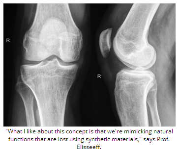
“What I like about this concept is that we’re mimicking natural functions that are lost using synthetic materials.”

Meanwhile in November 2013, Medical News Today learned how another team of researchers – also inspired by nature – is refining sea coral for use in bone grafts.

Written by Catharine Paddock PhD

http://www.medicalnewstoday.com/articles/281212.php


The American Academy of Ophthalmology offers guidance to help improve public understanding of cataracts

Ophthalmology

Cataract is one of the leading causes of blindness in the United States. Approximately 24.5 million Americans have the lens-clouding eye condition, and the incidence is set to grow by 50 percent by 2020.[i] As part of its efforts to support Cataract Awareness Month this June, the American Academy of Ophthalmology – the world’s largest association of eye physicians and surgeons – is sharing with the public hundreds of commonly-asked questions and answers about the condition, which affects more than half of all Americans by age 80.[ii]

If not treated through a change in eyeglass prescription or surgery, cataracts can increase risk of permanent blindness. In addition, the longer advanced cataracts are left untreated, the more difficult it can be to successfully remove the cataract and restore vision. To help people understand the condition, its causes and treatments, Academy member ophthalmologists – medical doctors specializing in the diagnosis, medical and surgical treatment of eye diseases and conditions – have responded to hundreds of queries about cataracts submitted by the public through the Ask an Eye M.D. portal onGetEyeSmart.org. The following five questions and answers about cataracts are a small sampling of what is available for public reference on the website:

Can you have 20/20 vision and still be diagnosed with cataract?

“Yes, you can,” answers Jeffrey Whitman, M.D. “Having a cataract just means that the lens of your eye has become cloudy and hardened – a process that begins at around 50 years of age and does not preclude 20/20 vision. It is only when it becomes visually significant – that is, when it degrades your vision, changes color perception, or causes glare at nighttime – that it requires surgical care.”

How can I keep cataracts from getting worse?

“Most individuals over age 50 to 60 technically have age-related changes in their lenses that might be termed ‘very early cataracts,'” says Charles P. Wilkinson, M.D. “In general, prevention is very difficult; but the most helpful practices include:

  • Avoid ultraviolet light from the sun with sunglasses
  • Avoid using steroid eye drops unless absolutely necessary
  • Avoid the rare medications that may be associated with cataract progression, including psoralens, a drug used along with light therapy to treat skin disorders; chlorpromazine, an antipsychotic; and someglaucoma medications.”

Do cataracts cause eye pain?

“Cataracts do not cause pain except if they have been allowed to remain untreated for too long,” explains Wayne Bizer, D.O. “In this case they cause a lot of pain and light sensitivity. Consult your ophthalmologist immediately if you are having eye pain.”

Why do I need to stop wearing my contact lenses before cataract surgery?

“Before cataract surgery, important measurements of the surface of your eye must be taken,” says W. Barry Lee, M.D. “Contact lenses alter the shape of the eye’s surface, which can make the measurements inaccurate and lead to poor vision after the surgery. The length of time you must not wear contact lenses prior to your cataract surgery varies depending on the type of contact lenses you wear, so listen to your ophthalmologist’s instructions carefully.”

How long is recovery time after cataract surgery?

“Typically, this should only take several days,” says Gary Hirshfield, M.D. “Of course, some issues may occur that require a longer recovery period, such as other eye conditions or rare surgery complications. Additionally, if both eyes need to be done and you are significantly near- or far-sighted, then there may be a period of time in between the surgery for each eye where the differences between the eyes may make your tasks difficult. Also, depending upon the surgical approach you may need a change in your eyeglass prescription which is usually done at about four weeks. However that can be accelerated to just several days provided you understand that the prescription may need to be revised in several weeks or months.”

“While cataracts are one of the most common eye conditions – especially for older adults – when and why to seek treatment and what kind can be a complex decision,” said Daniel J. Briceland, M.D., ophthalmologist and clinical spokesperson for the American Academy of Ophthalmology. “Some people wait too long before seeing a doctor about a suspected cataract, but they should really see an ophthalmologist for a comprehensive eye exam. Even if immediate treatment is not required, at least an ophthalmologist can confirm this and have a baseline from which to compare your vision if and when the cataract worsens later on.”

Seniors who have not had an eye exam in the last three years and for whom cost is a concern may qualify for EyeCare America, a public service program of the Foundation of the American Academy of Ophthalmology, which provides eye exams and care at no out-of-pocket cost for eligible seniors age 65 and older through its network of more than 6,000 volunteer ophthalmologists. Visit www.eyecareamerica.org to see if you or your loved ones are eligible.

See all 250 cataract-related questions and answers or submit your own question at www.geteyesmart.org/ask.

http://www.medicalnewstoday.com/releases/277794.php


Eye Health and Retinal Detachment

Retinal detachment is a very serious eye condition that happens when the retina separates from the tissue around it. Since the retina can’t work properly under these conditions, you could permanently lose vision if the detached retina isn’t repaired promptly.

Who’s at Risk for a Detached Retina?

You’re more likely to get a detached retina if you:

  • Are severely nearsighted
  • Have had an eye injury or cataract surgery
  • Have a family history of retinal detachment

You may have heard the words “retinal tear,” too. That’s not the same as a retinal detachment.

  • Retinal tears often happen first. If fluid from within the eye passes through a retinal tear, that can separate the retina from its underlying tissue — and that’s retinal detachment. Retinal detachment may also happen with no warning. That’s more likely in elderly or very nearsighted people.

If you are unsure about your risk of retinal detachment, talk to your eye doctor.

What Are the Symptoms of a Detached Retina?

A detached retina doesn’t hurt, so look for these symptoms:

  • Flashes of light
  • Seeing “floaters” (small flecks or threads)
  • Darkening of your peripheral (side) vision

If you notice any of those symptoms, contact your eye doctor immediately.

How Is a Detached Retina Diagnosed?

Your health care provider would give you an eye exam, which would include dilating your eyes. That lets them see if your retina is detached.

Early diagnosis is key to preventing vision loss from a detached retina.

How Is a Detached Retina Treated?

There are many ways to treat a detached retina. These include:

  • Laser (thermal) or freezing (cryopexy). Both of these approaches can repair a tear in the retina if it is diagnosed early enough. This procedure is often done in the doctor’s office.
  • Pneumatic retinopexy. This procedure can be used to treat retinal detachment if the tear is small and easy to close. A small gas bubble is injected into the eye (specifically, into the  the clear, gel-like substance between the lens and the retina), where it then rises and presses against the retina, closing the tear. A laser or cryopexy can then be used to seal the tear.
  • Scleral buckle. This treatment for retinal detachment involves surgically sewing a silicone band (buckle) around the white of the eye (called the sclera) to push the sclera toward the tear until the tear heals. This band is not visible and remains permanently attached. Thermal treatment may then be necessary to seal the tear.
  • Vitrectomy. This surgery for retinal detachment is used for large tears. During a vitrectomy, the doctor removes the vitreous (the clear, gel-like substance between eye’s lens and retina) and replaces it with a saline solution.

Can a Detached Retina Be Prevented?

Yes, in some cases.

Getting an eye exam can flag early changes in your eyes that you may not have noticed. Treating those changes can help.

You should get your eyes checked once a year, or more often if you have conditions such as diabetes that make you more likely to have eye disease. Regular eye exams are also important if you are very nearsighted, as nearsightedness makes retinal detachment more likely.

If you have diabetes or high blood pressure, keeping those conditions under control will help the blood vessels in your retina, which is good for your eyes.

Not sure how often you should get your eyes checked? Ask your eye doctor.

You should also use the appropriate eye protection for certain activities. For instance, you should wear sports goggles with polycarbonate lenses while playing racquetball or certain other sports. You may also need eye protection if you work with machines, chemicals, or tools at work or home.

http://www.webmd.com/eye-health/eye-health-retinal-detachment