Tag Archives: EYE CARE

Wearable computer gloves ‘help teach braille’

Ophthalmology

More than 39 million people around the world are blind. For many of these individuals, braille – a reading and writing system that utilizes a series of raised dots that represent letters, numbers and punctuation – is a valuable tool. Now, researchers from the Georgia Institute of Technology have developed a wearable computer glove that can teach braille, even when the user’s attention is on another activity.

The research team – including Thad Starner, a professor at Georgia Tech and a technical/lead manager on Google’s Project Glass – first created a technology-enhanced glove back in 2008, called Piano Touch. The glove could teach individuals how to play piano melodies in 45 minutes.

Their latest creation is an advancement on Piano Touch, which has been built around a process called passive haptic learning (PHL) – the idea that people can learn a skill unconsciously without devoting full attention to what they are learning.

“We’ve learned that people can acquire motor skills through vibrations without devoting active attention to their hands,” says Tharner.

According to the researchers, only 10% of blind people learn braille. They believe it is something that is largely neglected in schools and note that the system can also be difficult to learn as a person ages, when blindness is most common. But could this new wearable technology help with the braille learning process?

Putting the glove to the test

The team put the new glove to the test in order to see how well the technology could teach braille.

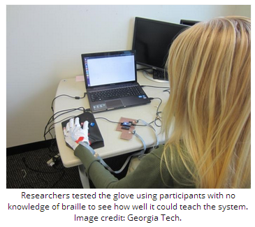
For their study, participants were required to wear the gloves during a series of tasks. The gloves consist of small vibrating motors that are stitched into the knuckles.

In the first task, the motors in the glove vibrated in a sequence that correlated with a typing pattern of a premeditated phrase in braille. The participants were given audio cues that let them know what braille letters were produced through typing that particular sequence.

Each participant was then required to type the phrase once on a keyboard without any vibrations or audio cues while the researchers measured their accuracy.

In the following task, participants were asked to play a computer game for 30 minutes – as a distraction – while wearing the glove. Half of the participants were presented with repeated vibrations and audio cues that represented the same braille phrase as the previous task, while the remaining participants acted as a control group and were only given audio cues.

The researchers note that the subjects had no previous knowledge of braille and the tasks did not include visual feedback, meaning participants were unaware of their accuracy.

Participants ‘could read and write braille’

On comparing the participants’ results with those of the first task, the team found that those in the control group had about the same level of accuracy.

However, those who had repeated vibrations and audio cues in the second task were a third more accurate, with some even gaining perfect accuracy. Furthermore, the researchers found that these participants were then able to effectively go from writing braille to reading it.

“After the typing test, passive learners were able to read and recognize more than 70% of the phrase’s letters,” says study co-author Caitlyn Seim, a student at Georgia Tech.

Seim is now in the process of conducting another study, which involves using the glove to teach the full braille alphabet to participants. She says that so far, 75% of subjects have demonstrated perfect typing accuracy. In addition, participants were able to recognize and read more than 90% of braille letters after 4 hours of learning.

Medical News Today recently reported on another creation for the visually impaired by researchers from Oxford University in the UK – “Smart glasses.” The glasses have been designed to help near-blind users navigate public spaces and better interact with others, as the glasses enhance facial features.

Written byHonor Whiteman

http://www.medicalnewstoday.com/articles/278719.php


The American Academy of Ophthalmology offers guidance to help improve public understanding of cataracts

Ophthalmology

Cataract is one of the leading causes of blindness in the United States. Approximately 24.5 million Americans have the lens-clouding eye condition, and the incidence is set to grow by 50 percent by 2020.[i] As part of its efforts to support Cataract Awareness Month this June, the American Academy of Ophthalmology – the world’s largest association of eye physicians and surgeons – is sharing with the public hundreds of commonly-asked questions and answers about the condition, which affects more than half of all Americans by age 80.[ii]

If not treated through a change in eyeglass prescription or surgery, cataracts can increase risk of permanent blindness. In addition, the longer advanced cataracts are left untreated, the more difficult it can be to successfully remove the cataract and restore vision. To help people understand the condition, its causes and treatments, Academy member ophthalmologists – medical doctors specializing in the diagnosis, medical and surgical treatment of eye diseases and conditions – have responded to hundreds of queries about cataracts submitted by the public through the Ask an Eye M.D. portal onGetEyeSmart.org. The following five questions and answers about cataracts are a small sampling of what is available for public reference on the website:

Can you have 20/20 vision and still be diagnosed with cataract?

“Yes, you can,” answers Jeffrey Whitman, M.D. “Having a cataract just means that the lens of your eye has become cloudy and hardened – a process that begins at around 50 years of age and does not preclude 20/20 vision. It is only when it becomes visually significant – that is, when it degrades your vision, changes color perception, or causes glare at nighttime – that it requires surgical care.”

How can I keep cataracts from getting worse?

“Most individuals over age 50 to 60 technically have age-related changes in their lenses that might be termed ‘very early cataracts,'” says Charles P. Wilkinson, M.D. “In general, prevention is very difficult; but the most helpful practices include:

  • Avoid ultraviolet light from the sun with sunglasses
  • Avoid using steroid eye drops unless absolutely necessary
  • Avoid the rare medications that may be associated with cataract progression, including psoralens, a drug used along with light therapy to treat skin disorders; chlorpromazine, an antipsychotic; and someglaucoma medications.”

Do cataracts cause eye pain?

“Cataracts do not cause pain except if they have been allowed to remain untreated for too long,” explains Wayne Bizer, D.O. “In this case they cause a lot of pain and light sensitivity. Consult your ophthalmologist immediately if you are having eye pain.”

Why do I need to stop wearing my contact lenses before cataract surgery?

“Before cataract surgery, important measurements of the surface of your eye must be taken,” says W. Barry Lee, M.D. “Contact lenses alter the shape of the eye’s surface, which can make the measurements inaccurate and lead to poor vision after the surgery. The length of time you must not wear contact lenses prior to your cataract surgery varies depending on the type of contact lenses you wear, so listen to your ophthalmologist’s instructions carefully.”

How long is recovery time after cataract surgery?

“Typically, this should only take several days,” says Gary Hirshfield, M.D. “Of course, some issues may occur that require a longer recovery period, such as other eye conditions or rare surgery complications. Additionally, if both eyes need to be done and you are significantly near- or far-sighted, then there may be a period of time in between the surgery for each eye where the differences between the eyes may make your tasks difficult. Also, depending upon the surgical approach you may need a change in your eyeglass prescription which is usually done at about four weeks. However that can be accelerated to just several days provided you understand that the prescription may need to be revised in several weeks or months.”

“While cataracts are one of the most common eye conditions – especially for older adults – when and why to seek treatment and what kind can be a complex decision,” said Daniel J. Briceland, M.D., ophthalmologist and clinical spokesperson for the American Academy of Ophthalmology. “Some people wait too long before seeing a doctor about a suspected cataract, but they should really see an ophthalmologist for a comprehensive eye exam. Even if immediate treatment is not required, at least an ophthalmologist can confirm this and have a baseline from which to compare your vision if and when the cataract worsens later on.”

Seniors who have not had an eye exam in the last three years and for whom cost is a concern may qualify for EyeCare America, a public service program of the Foundation of the American Academy of Ophthalmology, which provides eye exams and care at no out-of-pocket cost for eligible seniors age 65 and older through its network of more than 6,000 volunteer ophthalmologists. Visit www.eyecareamerica.org to see if you or your loved ones are eligible.

See all 250 cataract-related questions and answers or submit your own question at www.geteyesmart.org/ask.

http://www.medicalnewstoday.com/releases/277794.php


What Is Glaucoma? What Causes Glaucoma?

April_Part 1_Ophthalmology

Glaucoma is a disease of the eye in which fluid pressure within the eye rises – if left untreated the patient may lose vision, and even become blind. The disease generally affects both eyes, although one may have more severe signs and symptoms than the other.


There is a small space in the front of the eye called the “anterior chamber”. Clear liquid flows in-and-out of the anterior chamber, this fluid nourishes and bathes nearby tissues. If a patient has glaucoma, the fluid does not drain properly – it drains too slowly – out of the eye. This leads to fluid build-up, and pressure inside the eye rises. Unless this pressure is brought down and controlled, the optic nerve and other parts of the eye may become damaged, leading to loss of vision.


There are two main types of glaucoma, open angle andclosed angle (angle closure) glaucoma. The fluid in the eye flows through an area between the iris and cornea, where it escapes via the trabecular meshwork – “angle” refers to this area. The trabecular meshwork is made of sponky tissue lined by trabeculocytes. Fluid drains into s set of tubes, known as Schlemm’s canal, from which they flow into the blood system.


Closed Angle Glaucoma (acute angle-closure glaucoma) can come on suddenly, and the patient commonly experiences pain and rapid vision loss. Fortunately, the symptoms of pain and discomfort make the sufferer seek medical help, resulting in prompt treatment which usually prevents any permanent damage from occurring.


Primary Open Angle Glaucoma (chronic glaucoma) – progresses very slowly. The patient may not feel any symptoms; even slight loss of vision may go unnoticed. In this type of glaucoma, many people don’t get medical help until some permanent damage has already occurred.


Low-tension glaucoma – this is another form that experts do not fully understand. Even though eye pressure is normal, optic nerve damage still occurs. Perhaps the optic nerve is over-sensitive or there is atherosclerosisin the blood vessel that supplies the optic nerve.


Pigmentary glaucoma – this type generally develops during early or middle adulthood. Pigment granules, which arise from the back of the iris, are dispersed within the eye. If these granules build up in the trabecular meshwork, they can undermine the flow of fluids in the eye, leading to a rise in eye pressure. Running and some other sports can unsettle the granules, which get into the travecular meshwork.

Glaucoma has been called the silent thief of sight


Primary glaucoma – this means we do not know what the cause was.


Secondary glaucoma – the condition has a known cause, such as atumor, diabetes, an advanced cataract, or inflammation.

What are the signs and symptoms of glaucoma?

A symptom is something the sufferer experiences and describes, such as pain, while a sign is something others can identify, such as a rash or a swelling.


The signs and symptoms of primary open angle glaucoma and acute angle-closure glaucoma are quite different.


Signs and symptoms of primary open-angle glaucoma

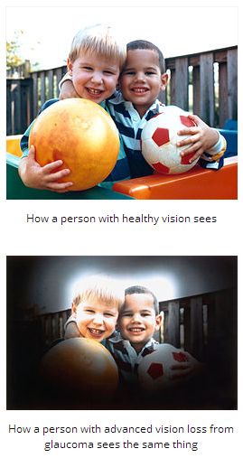
  • Peripheral vision is gradually lost. This nearly always affects both eyes.
  • In advanced stages, the patient has tunnel vision

Signs and symptoms of closed angle glaucoma

  • Eye pain, usually severe
  • Blurred vision
  • Eye pain is often accompanied by nausea, and sometimes vomiting
  • Lights appear to have extra halo-like glows around them
  • Red eyes
  • Sudden, unexpected vision problems, especially when lighting is poor

What risk factors are linked to glaucoma?

A risk factor is something that raises the risk of developing a condition or disease. For example, obesity is a risk factor for diabetes type 2 – obese people have a higher risk of developing diabetes.

  • Old age – people over the age of 60 years have a higher risk of developing the disease. For African-Americans, the risk rises at a younger age.
  • Ethnic background – East Asians, because of their shallower anterior chamber depth, have a higher risk of developing glaucoma compared to Caucasians. The risk for those of Inuit origin is considerably greater still. People of African-American descent are three to four times more likely to develop the disease compared to American whites. Females are three times as likely to develop glaucoma as males.
  • Some illnesses and conditions – people with diabetes or hypothyroidism have a much higher chance of developing glaucoma.
  • Eye injuries or conditions – some eye injuries, especially severe ones, are linked to a higher glaucoma risk.Retinal detachment, eye inflammations and eye tumors can also cause glaucoma to occur.
  • Eye surgery – some patients who underwent eye surgery have a higher risk of glaucoma.
  • Myopia – people with myopia (nearsightedness) have a higher risk of glaucoma.
  • Corticosteroids – patients on long-term corticosteroids have a raised risk of developing several different conditions, including glaucoma. The risk is even greater with eyedrops containing corticosteroids.

Diagnosing glaucoma

Eye-pressure test – the doctor uses a tonometer, a device which measures intraocular pressure (pressure inside the eye). Some anesthetic and a dye is placed in the cornea, and a blue light is held against the eye to measure pressure. This test can diagnose ocular hypertension; a risk factor for open-angle glaucoma.


The doctor also measures corneal thickness, because it affects how the pressure inside the eye is interpreted.


Gonioscopy – this examines the area where the fluid drains out of the eye. It helps determine whether the angle between the cornea and the iris is open or blocked (closed).


Perimetry test – also known as a visual field test. It determines which area of the patient’s vision is missing. The patient is shown a sequence of light spots and asked to identify them. Some of the dots are located where the person’s peripheral vision is; the part of vision that is initially affected by glaucoma. If the patient cannot see those peripheral dots, it means that some vision damage has already occurred.


Optic nerve damage – the ophthalmologist (eye doctor) uses instruments to look at the back of the eye, which can reveal any slight changes which may also point towards glaucoma onset.

What are the treatment options for glaucoma?

Treatments involve either improving the flow of fluid inside the eye, reducing its production, and sometimes both. Damage caused by glaucoma is irreversible. Even the disease itself cannot be completely cured. However, regular check-ups and proper treatment can considerably slow down the progression of the disease, and even prevent further loss of eyesight.


Eyedrops – in the majority of cases, initial treatment includes eyedrops. Compliance is vital for best results and to prevent undesirable side effects – this means following the doctor’s instructions carefully. Examples of eyedrops include:

  • Prostaglandin analogues – these medications have prostaglandin-like compounds as their active ingredient. They increase the outflow of the fluid inside the eye. Some patients may experience reddening and stinging of the eyes, photophobia, some swelling around the rim of the eye, as well as darkening of the iris. The color of the eyelids may also change, and there may be blurred vision. Examples include Xalatan (latanoprost) and Lumigan (bimatroprost).
  • Beta blockers – these medication reduce the amount of fluid the eye produces. Some patients may experience breathing problems, hair loss, fatigue, depression, memory loss, a drop in blood pressure, and/or impotence. Examples of such medications include timolol, betaxolol and metipranolol.
  • Patients with certain lung conditions, such as emphysema or bronchitis may be prescribed a different medication. Diabetes patients who are taking insulin may also be prescribed an alternative drug.
  • Carbonic anhydrase inhibitors – these also reduce fluid production in the eye. Side effects may include nausea, eye irritation, dry mouth, frequent urination, tingling in the fingers/toes, and a strange taste in the mouth. Examples include brinzolamide and dorzolamide.
  • Cholinergic agents – also known as miotic agents. They help the fluids flow out of the eye. Side effects may include pain in and around the eye, myopia, blocked nose, some digestive problems, increased sweating and/or salivation, blurred vision, and ache around the eyebrows. Examples include pilocarpine and carbachol.

Sympathomimetic drugs – these reduce the production of fluid in the eye, as well as increasing their flow out of the eye. An example is dipivefrin, an epinephrine (adrenaline) compound. Some patients may experience painful and red eyes. Make sure your doctor knows if you suffer from heart disease or hypertension (high blood pressure).If eyedrops are not effective enough, the doctor may prescribe an oral carbonic anhydrase inhibitor. Side effects are less if they are taken during meals. Initial side effects may include tingling in the fingers and toes and frequent urination – however, after a few days they usually resolve. Much less commonly, there is also a risk of rashes, kidney stones, stomachache, weight loss, impotence, fatigue, and a strange taste when consuming fizzy drinks.


Surgery – if drugs don’t work, or if the patient cannot tolerate them, surgical intervention may become an option. The aim of surgery is usually to bring down the pressure inside the eye. Examples of surgery may include:

  • Trabeculoplasty – a high-energy laser beam is used to unblock clogged drainage canals, making it easier for the fluid inside the eye to drain out. This procedure nearly always reduces inner eye pressure. However, the problem may come back.
  • Filtering surgery (viscocanalostomy) – if nothing else works, including high-beam energy laser surgery, the patient may need a filtering procedure, usually a type of trabeculectomy. The surgeon creates an opening in the white of the eye and removes a small piece of the trabecular meshwork. This allows the eye fluid to leave through the opening.
  • Drainage implant (aqueous shunt implant) – this option is sometimes used for children or those with secondary glaucoma. A small silicone tube is inserted into the eye to help it drain out fluids better.

Acute angle-closure glaucoma – this condition is treated as a medical emergency. Pressure-reducing medications are administered immediately. A laser procedure is usually carried out which creates a tiny hole in the iris, allowing fluids to pass into the trabecular meshwork – this procedure is called an iridotomy. Even if only one eye is affected, the doctor may decide to treat both, because this type of glaucoma often affects the other eye too.

Complications of glaucoma

Loss of eyesight is the most common complication. The earlier a patient is diagnosed with glaucoma, the better his/her outlook.


Written by Christian Nordqvist

http://www.medicalnewstoday.com/articles/9710.php
What Is Glaucoma? What Causes Glaucoma?

Glaucoma is a disease of the eye in which fluid pressure within the eye rises – if left untreated the patient may lose vision, and even become blind. The disease generally affects both eyes, although one may have more severe signs and symptoms than the other.


There is a small space in the front of the eye called the “anterior chamber”. Clear liquid flows in-and-out of the anterior chamber, this fluid nourishes and bathes nearby tissues. If a patient has glaucoma, the fluid does not drain properly – it drains too slowly – out of the eye. This leads to fluid build-up, and pressure inside the eye rises. Unless this pressure is brought down and controlled, the optic nerve and other parts of the eye may become damaged, leading to loss of vision.


There are two main types of glaucoma, open angle andclosed angle (angle closure) glaucoma. The fluid in the eye flows through an area between the iris and cornea, where it escapes via the trabecular meshwork – “angle” refers to this area. The trabecular meshwork is made of sponky tissue lined by trabeculocytes. Fluid drains into s set of tubes, known as Schlemm’s canal, from which they flow into the blood system.


Closed Angle Glaucoma (acute angle-closure glaucoma) can come on suddenly, and the patient commonly experiences pain and rapid vision loss. Fortunately, the symptoms of pain and discomfort make the sufferer seek medical help, resulting in prompt treatment which usually prevents any permanent damage from occurring.


Primary Open Angle Glaucoma (chronic glaucoma) – progresses very slowly. The patient may not feel any symptoms; even slight loss of vision may go unnoticed. In this type of glaucoma, many people don’t get medical help until some permanent damage has already occurred.


Low-tension glaucoma – this is another form that experts do not fully understand. Even though eye pressure is normal, optic nerve damage still occurs. Perhaps the optic nerve is over-sensitive or there is atherosclerosisin the blood vessel that supplies the optic nerve.


Pigmentary glaucoma – this type generally develops during early or middle adulthood. Pigment granules, which arise from the back of the iris, are dispersed within the eye. If these granules build up in the trabecular meshwork, they can undermine the flow of fluids in the eye, leading to a rise in eye pressure. Running and some other sports can unsettle the granules, which get into the travecular meshwork.

Glaucoma has been called the silent thief of sight


Primary glaucoma – this means we do not know what the cause was.


Secondary glaucoma – the condition has a known cause, such as atumor, diabetes, an advanced cataract, or inflammation.

What are the signs and symptoms of glaucoma?

A symptom is something the sufferer experiences and describes, such as pain, while a sign is something others can identify, such as a rash or a swelling.


The signs and symptoms of primary open angle glaucoma and acute angle-closure glaucoma are quite different.


Signs and symptoms of primary open-angle glaucoma

  • Peripheral vision is gradually lost. This nearly always affects both eyes.
  • In advanced stages, the patient has tunnel vision

Signs and symptoms of closed angle glaucoma

  • Eye pain, usually severe
  • Blurred vision
  • Eye pain is often accompanied by nausea, and sometimes vomiting
  • Lights appear to have extra halo-like glows around them
  • Red eyes
  • Sudden, unexpected vision problems, especially when lighting is poor

What risk factors are linked to glaucoma?

A risk factor is something that raises the risk of developing a condition or disease. For example, obesity is a risk factor for diabetes type 2 – obese people have a higher risk of developing diabetes.

  • Old age – people over the age of 60 years have a higher risk of developing the disease. For African-Americans, the risk rises at a younger age.
  • Ethnic background – East Asians, because of their shallower anterior chamber depth, have a higher risk of developing glaucoma compared to Caucasians. The risk for those of Inuit origin is considerably greater still. People of African-American descent are three to four times more likely to develop the disease compared to American whites. Females are three times as likely to develop glaucoma as males.
  • Some illnesses and conditions – people with diabetes or hypothyroidism have a much higher chance of developing glaucoma.
  • Eye injuries or conditions – some eye injuries, especially severe ones, are linked to a higher glaucoma risk.Retinal detachment, eye inflammations and eye tumors can also cause glaucoma to occur.
  • Eye surgery – some patients who underwent eye surgery have a higher risk of glaucoma.
  • Myopia – people with myopia (nearsightedness) have a higher risk of glaucoma.
  • Corticosteroids – patients on long-term corticosteroids have a raised risk of developing several different conditions, including glaucoma. The risk is even greater with eyedrops containing corticosteroids.

Diagnosing glaucoma

Eye-pressure test – the doctor uses a tonometer, a device which measures intraocular pressure (pressure inside the eye). Some anesthetic and a dye is placed in the cornea, and a blue light is held against the eye to measure pressure. This test can diagnose ocular hypertension; a risk factor for open-angle glaucoma.


The doctor also measures corneal thickness, because it affects how the pressure inside the eye is interpreted.


Gonioscopy – this examines the area where the fluid drains out of the eye. It helps determine whether the angle between the cornea and the iris is open or blocked (closed).


Perimetry test – also known as a visual field test. It determines which area of the patient’s vision is missing. The patient is shown a sequence of light spots and asked to identify them. Some of the dots are located where the person’s peripheral vision is; the part of vision that is initially affected by glaucoma. If the patient cannot see those peripheral dots, it means that some vision damage has already occurred.


Optic nerve damage – the ophthalmologist (eye doctor) uses instruments to look at the back of the eye, which can reveal any slight changes which may also point towards glaucoma onset.

What are the treatment options for glaucoma?

Treatments involve either improving the flow of fluid inside the eye, reducing its production, and sometimes both. Damage caused by glaucoma is irreversible. Even the disease itself cannot be completely cured. However, regular check-ups and proper treatment can considerably slow down the progression of the disease, and even prevent further loss of eyesight.


Eyedrops – in the majority of cases, initial treatment includes eyedrops. Compliance is vital for best results and to prevent undesirable side effects – this means following the doctor’s instructions carefully. Examples of eyedrops include:

  • Prostaglandin analogues – these medications have prostaglandin-like compounds as their active ingredient. They increase the outflow of the fluid inside the eye. Some patients may experience reddening and stinging of the eyes, photophobia, some swelling around the rim of the eye, as well as darkening of the iris. The color of the eyelids may also change, and there may be blurred vision. Examples include Xalatan (latanoprost) and Lumigan (bimatroprost).
  • Beta blockers – these medication reduce the amount of fluid the eye produces. Some patients may experience breathing problems, hair loss, fatigue, depression, memory loss, a drop in blood pressure, and/or impotence. Examples of such medications include timolol, betaxolol and metipranolol.
  • Patients with certain lung conditions, such as emphysema or bronchitis may be prescribed a different medication. Diabetes patients who are taking insulin may also be prescribed an alternative drug.
  • Carbonic anhydrase inhibitors – these also reduce fluid production in the eye. Side effects may include nausea, eye irritation, dry mouth, frequent urination, tingling in the fingers/toes, and a strange taste in the mouth. Examples include brinzolamide and dorzolamide.
  • Cholinergic agents – also known as miotic agents. They help the fluids flow out of the eye. Side effects may include pain in and around the eye, myopia, blocked nose, some digestive problems, increased sweating and/or salivation, blurred vision, and ache around the eyebrows. Examples include pilocarpine and carbachol.

Sympathomimetic drugs – these reduce the production of fluid in the eye, as well as increasing their flow out of the eye. An example is dipivefrin, an epinephrine (adrenaline) compound. Some patients may experience painful and red eyes. Make sure your doctor knows if you suffer from heart disease or hypertension (high blood pressure).If eyedrops are not effective enough, the doctor may prescribe an oral carbonic anhydrase inhibitor. Side effects are less if they are taken during meals. Initial side effects may include tingling in the fingers and toes and frequent urination – however, after a few days they usually resolve. Much less commonly, there is also a risk of rashes, kidney stones, stomachache, weight loss, impotence, fatigue, and a strange taste when consuming fizzy drinks.


Surgery – if drugs don’t work, or if the patient cannot tolerate them, surgical intervention may become an option. The aim of surgery is usually to bring down the pressure inside the eye. Examples of surgery may include:

  • Trabeculoplasty – a high-energy laser beam is used to unblock clogged drainage canals, making it easier for the fluid inside the eye to drain out. This procedure nearly always reduces inner eye pressure. However, the problem may come back.
  • Filtering surgery (viscocanalostomy) – if nothing else works, including high-beam energy laser surgery, the patient may need a filtering procedure, usually a type of trabeculectomy. The surgeon creates an opening in the white of the eye and removes a small piece of the trabecular meshwork. This allows the eye fluid to leave through the opening.
  • Drainage implant (aqueous shunt implant) – this option is sometimes used for children or those with secondary glaucoma. A small silicone tube is inserted into the eye to help it drain out fluids better.

Acute angle-closure glaucoma – this condition is treated as a medical emergency. Pressure-reducing medications are administered immediately. A laser procedure is usually carried out which creates a tiny hole in the iris, allowing fluids to pass into the trabecular meshwork – this procedure is called an iridotomy. Even if only one eye is affected, the doctor may decide to treat both, because this type of glaucoma often affects the other eye too.

Complications of glaucoma

Loss of eyesight is the most common complication. The earlier a patient is diagnosed with glaucoma, the better his/her outlook.


Written by Christian Nordqvist

http://www.medicalnewstoday.com/articles/9710.php


Bevacizumab – Treatment For Diabetic Macular Edema

Ophthalmology

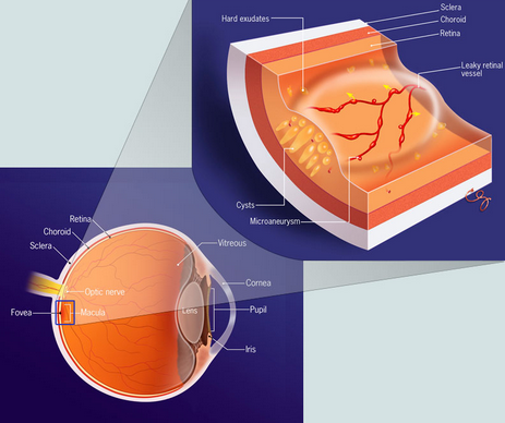
According to a study published Online First by Archives of Ophthalmology, bevacizumab appears to be more effective at treating diabetic macular edema (swelling of the retina) thanmacular laser therapy.


The researchers of the randomized controlled trial found that among participants with persistent clinically significant diabetic macular edema (CSME), bevacizumab showed to be effective at 12 months and was maintained through 24 months.


For the past 30 years, Modified Early Treatment Diabetic Retinopathy Study (ETDRS) macular laser therapy (MLT) has been the leading treatment for individuals suffering from CSME.


Although MLT lowers the risk of moderate visual loss, improved treatments have been sought as visual acuity only improves in less than 3% of patients (a 15-letter gain at 3 years).


Ranjan Rajendram, M.D., F.R.C.Ophth., of Moorfields Eye Hospital, London, and colleagues reveal the two-year outcomes of the BOLT Study.


The prospective randomized controlled trial involved 80 participants with CSME. Participants were randomly assigned to receive either injections into the eye (intravitreous bevacizumab) or modified ETDRS MLT.


The researchers found that at two years, the mean ETDRS best-corrected visual acuity in the bevacizumab group was 64.4 (Snellen visual acuity equivalent: 20/50) and 54.8 (Snellen equivalent: 20/80) in the MLT group.


Participants in the bevacizumab group gained a mean 8.6 letters, while participants in the MLT group lost a mean of 0.5 letters.


At 24 months, patients in the bevacizumab group gained a median of 9 ETDRS letters, compared with 2.5 ETDRS letters in the MLT group.


At two years, 49% of patients in the bevacizumab group gained 10+ ETDRS letters and 32 gained 15+ letters, versus participants in the MLT group 7% and 4%.


The researchers conclude:


“In conclusion, this investigator-initiated single-center study provides evidence for the longer-term use of bevacizumab in the treatment of persistent DME [diabetic macular edema].


Visual acuity benefit was maintained through two years with a reduced injection frequency in the second year despite the long duration of DME and multiple MLTs before entering the study. This finding will be reassuring to physicians charged with delivery of this relatively new treatment.”


Written By Grace Rattue


http://www.medicalnewstoday.com/articles/244008.php

Picture courtesy of www.treatmydme.com


Stop Smoking, Cut Cataract Risk

Ophthalmology

The cataract risk associated with smoking slowly fell after ceasing tobacco use, although not to the level of a never-smoker, a Swedish population-based study showed.

Smoking more than 15 cigarettes per day was associated with 42% higher likelihood of a cataract surgery during 12 years of follow-up, Birgitta Ejdervik Lindblad, MD, PhD, of Örebro University Hospital in Örebro, Sweden, and colleagues found.

After having quit for 20 years, that risk had declined to a relative 21% above that of never-smokers, the researchers reported online in JAMA Ophthalmology.

The risk declined significantly with time (P<0.001), but “the higher the intensity of smoking, the longer it takes for the increased risk to decline,” the researchers noted.

Even lighter smokers with a fewer-than-15-a-day habit remained at significant risk 2 decades after quitting (rate ratio 1.13, 95% CI 1.04-1.24).

“These findings emphasize the importance of early smoking cessation and preferably the avoidance of smoking,” Linblad’s group concluded.

Plenty of prior studies have shown cataract and other ocular risks from smoking, so “eye care professionals should encourage people to stop smoking,” too, they recommended.

The presumed mechanism for this risk is that “smoking increases the oxidative stress in the lens by generating free radicals and reduces the plasma concentration of several antioxidants, such as ascorbic acid,” Lindblad and colleagues explained.

“Cigarette smoke also contains toxic metal ions, and cadmium can accumulate in cataractous lenses of smokers,” they added. “Cadmium may affect anti-oxidative lens enzymes such as superoxide dismutase and glutathione peroxidase, thereby weakening the defense against oxidative damage and hastening cataract development.”

Their analysis included 44,371 men in the Cohort of Swedish Men study ages 45 to 79 years, among whom 25% reported smoking and 39% had been smokers, based on questionnaire responses regarding smoking habits and lifestyle factors.

During 12 years of follow-up via the Swedish National Day-Surgery Register, and local registers of cataract extraction in the study area, an age-adjusted 13% had cataract extractions.

Limitations of the study included possible misclassification of self-reported smoking history, no assessment or control for sunlight or UV exposure, and lack of data on subtype of cataracts, although all were severe enough to cause visual impairment requiring lens extraction, “and therefore having the greatest clinical and public health importance.”

The study was supported by grants from the Swedish Council for Working Life and Social Research and the Örebro County Council.

The researchers reported no conflicts of interest.

http://www.medpagetoday.com/PrimaryCare/Smoking/43653


Eye Health and Retinal Detachment

Retinal detachment is a very serious eye condition that happens when the retina separates from the tissue around it. Since the retina can’t work properly under these conditions, you could permanently lose vision if the detached retina isn’t repaired promptly.

Who’s at Risk for a Detached Retina?

You’re more likely to get a detached retina if you:

  • Are severely nearsighted
  • Have had an eye injury or cataract surgery
  • Have a family history of retinal detachment

You may have heard the words “retinal tear,” too. That’s not the same as a retinal detachment.

  • Retinal tears often happen first. If fluid from within the eye passes through a retinal tear, that can separate the retina from its underlying tissue — and that’s retinal detachment. Retinal detachment may also happen with no warning. That’s more likely in elderly or very nearsighted people.

If you are unsure about your risk of retinal detachment, talk to your eye doctor.

What Are the Symptoms of a Detached Retina?

A detached retina doesn’t hurt, so look for these symptoms:

  • Flashes of light
  • Seeing “floaters” (small flecks or threads)
  • Darkening of your peripheral (side) vision

If you notice any of those symptoms, contact your eye doctor immediately.

How Is a Detached Retina Diagnosed?

Your health care provider would give you an eye exam, which would include dilating your eyes. That lets them see if your retina is detached.

Early diagnosis is key to preventing vision loss from a detached retina.

How Is a Detached Retina Treated?

There are many ways to treat a detached retina. These include:

  • Laser (thermal) or freezing (cryopexy). Both of these approaches can repair a tear in the retina if it is diagnosed early enough. This procedure is often done in the doctor’s office.
  • Pneumatic retinopexy. This procedure can be used to treat retinal detachment if the tear is small and easy to close. A small gas bubble is injected into the eye (specifically, into the  the clear, gel-like substance between the lens and the retina), where it then rises and presses against the retina, closing the tear. A laser or cryopexy can then be used to seal the tear.
  • Scleral buckle. This treatment for retinal detachment involves surgically sewing a silicone band (buckle) around the white of the eye (called the sclera) to push the sclera toward the tear until the tear heals. This band is not visible and remains permanently attached. Thermal treatment may then be necessary to seal the tear.
  • Vitrectomy. This surgery for retinal detachment is used for large tears. During a vitrectomy, the doctor removes the vitreous (the clear, gel-like substance between eye’s lens and retina) and replaces it with a saline solution.

Can a Detached Retina Be Prevented?

Yes, in some cases.

Getting an eye exam can flag early changes in your eyes that you may not have noticed. Treating those changes can help.

You should get your eyes checked once a year, or more often if you have conditions such as diabetes that make you more likely to have eye disease. Regular eye exams are also important if you are very nearsighted, as nearsightedness makes retinal detachment more likely.

If you have diabetes or high blood pressure, keeping those conditions under control will help the blood vessels in your retina, which is good for your eyes.

Not sure how often you should get your eyes checked? Ask your eye doctor.

You should also use the appropriate eye protection for certain activities. For instance, you should wear sports goggles with polycarbonate lenses while playing racquetball or certain other sports. You may also need eye protection if you work with machines, chemicals, or tools at work or home.

http://www.webmd.com/eye-health/eye-health-retinal-detachment

Computer Use and Eye Strain

Staring at your computer screen, smartphone, video game or other digital devices for long periods won’t cause permanent eye damage, but your eyes may feel dry and tired. Some people also experience headaches or motion sickness when viewing 3-D, which may indicate that the viewer has a problem with focusing or depth perception.

What causes computer-use eyestrain?

Woman looking at laptop
  • Normally, humans blink about 18 times a minute, but studies show we blink half that often while using computers and other digital screen devices, whether for work or play.
  • Extended reading, writing or other intensive “near work” can also cause eyestrain.

What to do:

  • Sit about 25 inches from the computer screen and position the screen so your eye gaze is slightly downward.
  • Reduce glare from the screen by lighting the area properly; use a screen filter if needed.
  • Post a note that says “Blink!” on the computer as a reminder.
  • Every 20 minutes, shift your eyes to look at an object at least 20 feet away, for at least 20 seconds: the “20-20-20” rule.
  • Use artificial tears to refresh your eyes when they feel dry.
  • Take regular breaks from computer work, and try to get enough sleep at night.

Computer-use eyestrain can be made worse by:

Sleep deprivation. When you get less sleep than you need, your eyes may become irritated. During sleep our eyes rest for an extended period and are replenished by nutrients. Ongoing eye irritation can lead to swelling and infection, especially if you wear contact lenses.

  • If you have to be at your computer for a marathon work session, take regular rest breaks or “power naps,” if possible.
  • Apply a washcloth soaked in warm water to tired, dry eyes (with eyes closed).
  • Use tired or sore eyes as a signal that it’s time to stop working and get some rest or sleep.

Incorrect contact lens use. If you wear contact lenses, it’s important that you use and care for them properly — especially if you use a computer and other digital-screen devices often. This helps avoid eye irritation, swelling, infection and vision problems.

  • Give your eyes a break: wear your glasses!
  • Don’t sleep in your contact lenses, even if they are labeled “extended wear.”
  • Always use good cleaning practices.

There are some important things to keep in mind when cleaning your contact lenses. You should:

  • Avoid touching the lenses with water; use fresh solution every time for cleaning and storing.
  • Rub your contacts when you clean them, even if you use a no-rub solution.
  • Clean your storage case regularly (with fresh solution, not water) and replace it every 2 to 3 months.

Stop wearing your contact lenses and see an ophthalmologist (Eye M.D.) right away if you develop any of these problems: Eyes that are red, blurry, watery, sensitive to light, or sore; eye swelling or discharge.

http://www.geteyesmart.org/eyesmart/living/computer-usage.cfm

Myths and Facts About Ophthalmology

Myth

Reading in dim light is harmful to your eyes.

Fact

Although reading in dim light can make your eyes feel tired, it is not harmful.

Myth

It is not harmful to watch a welder or look at the sun if you squint, or look through narrowed eyelids.

Fact

Even if you squint, ultra-violet light still gets to your eyes, damaging the cornea, lens, and retina. Never watch welding without wearing the proper protection. Never look directly at an eclipse.

Myth

Using a computer, or video display terminal (VDT), is harmful to the eyes.

Fact

Although using a VDT is associated with eyestrain or fatigue, it is not harmful to the eyes.

Myth

If you use your eyes too much, you wear them out.

Fact

You can use your eyes as much as you wish – they do not wear out.

Myth

Wearing poorly-fit glasses damages your eyes.

Fact

Although a good fit is required for good vision with glasses, a poor fit does not damage your eyes.

Myth

Wearing poorly fit contacts does not harm your eyes.

Fact

Poorly fit contact lenses can be harmful to your cornea (the window at the front of your eye). Make certain your eyes are checked regularly by an eye doctor at our ophthalmology office if you wear contact lenses.

Myth

You do not need to have your eyes checked until you are in your 40s or 50s.

Fact

There are several asymptomatic, yet treatable, eye diseases (most notably glaucoma) that can begin prior to your 40s.

Myth

Safety goggles are more trouble than they’re worth.

Fact

Safety goggles prevent many potentially blinding injuries every year. Keep goggles handy and use them.

Myth

It’s okay to swim while wearing soft contact lenses.

Fact

Potentially blinding eye infections can result from swimming or using a hot tub while wearing contact lenses.

Myth

Children outgrow crossed eyes.

Fact

Children do not outgrow truly crossed eyes. A child whose eyes are misaligned has strabismus and can develop poor vision in one eye (a condition known as amblyopia) because the brain turns off the misaligned or “lazy” eye. The sooner crossed or misaligned eyes are treated, the less likely the child will have permanently impaired vision.

Myth

A cataract must be ripe before it can be removed.

Fact

With modern cataract surgery, a cataract does not have to ripen before it is removed. When a cataract keeps you from doing the things you like or need to do, consider having it removed.

Myth

Cataracts can be removed with lasers.

Fact

A cataract cannot be removed with a laser. The cloudy lens must be removed through a surgical incision. However, after cataract surgery, a membrane within the eye may become cloudy. This membrane can be opened with laser eye surgery.

Myth

Eyes can be transplanted.

Fact

The eye cannot be transplanted. It is connected to the brain by the optic nerve, which cannot be reconnected once it has been severed. A person can, however, undergo cornea transplant surgery to replace the cornea, the clear part of the eye. Surgeons often use plastic intraocular lens implants (IOLs) such as crystalens®, ReSTOR®, ReZoom™ or TECNIS® to replace natural lenses during cataract surgery.

http://www.batravision.com/html/eye-care.html

Link


glasses-jpgBy Pure Matters

Imagine what life would be like if you couldn’t see well. Reading might be out. Watching a movie could be tough. Focusing on the face of a loved one could drive you to tears.

The number of people losing their vision is growing, yet experts say much of this vision loss could be prevented.

“We can intervene best when we identify a problem in the early stages,” says Roy S. Rubinfeld, M.D., a clinical correspondent for the American Academy of Ophthalmology (AAO). He warns against putting off regular eye exams because your eyes feel fine or you don’t wear glasses or contact lenses. Signs of some eye diseases, such as glaucoma and age-related macular degeneration (AMD), are present before you might notice symptoms.


Growing number


The National Eye Institute says more than 3.3 million Americans ages 40 and older have blindness or low vision. The institute projects that figure will increase markedly by the year 2020. The percentage of people more than 60 years old who suffer vision loss is growing fast, too.


“At 60, everyone should have an annual eye exam, even if you’re seeing very well,” Dr. Rubinfeld says.


Many diseases cause vision loss as we age, but AMD is the Western world’s top cause of blindness. Leading to loss of your central vision, it may cause dark spots in your sight, make straight lines appear wavy, or cause text to seem blurry. AMD, glaucoma, diabetic retinopathy, cataracts, and dry eye syndrome can all rob you of sight.


It’s best to see your eye doctor before trouble starts. But these signs should prompt a visit at once:


  • Trouble seeing objects close up or far away
  • Colors that seem faded
  • Poor night vision
  • Double or multiple vision
  • Loss of side vision
  • Poor central vision or straight objects that look wavy
  • Blurry text or type


Save aging eyes


Dr. Rubinfeld offers these recommendations: