Author Archives: drreneebovelle

New Layer In Human Eye Discovered

A new layer in the front layer of the human eye has been discovered by researchers at The University of Nottingham.

The findings, published in the journal Ophthalmology, could significantly help doctors carry out corneal grafts or transplants.

The layer has been called the “Dua’s Layer”, named after the researcher who led the study, Professor Harminder Dua.

Harminder Dua, Professor of Ophthalmology and Visual Sciences, said that the discovery means that ophthalmology textbooks will literally have to be re-written.

He added:

“Having identified this new and distinct layer deep in the tissue of the cornea, we can now exploit its presence to make operations much safer and simpler for patients.”

Clinicians across the world are starting to relate the tear or absence in this layer to diseases at the back of the cornea.

The cornea is located on the front of the eye and allows light to enter. Previously believed for made up of five different layers:

  • Bowman’s layer
  • The corneal epithelium
  • The corneal stroma
  • Descemet’s membrane
  • The cornel endothelium

The Dua’s layer is located in the back of the cornea between the corneal stroma and Descemet’s membrane. It is extremely tough and strong despite being only 15 microns thick, it also impervious to air.

Iris - left eye of a girl
The newly discovered layer of the cornea is a significant advancement in our understanding of the human eye.

The new layer was discovered by simulating human corneal transplants on eyes collected from donors across the UK – given recent success in cultivated stem cells on human corneas, there might not be a need for donors in the future.

Small air bubbles were injected into the cornea of the eye to separate it into different layers. The researchers were able to study the layers a thousand times their actual size with electron microscopy.

Surgeons will benefit considerably by understanding more about the new Dua’s layer, which will improve outcomes for patients undergoing corneal grafts and transplants. There are over 65,000 penetrating corneal graft procedures being carried out worldwide each year, according to Eye Journal.

During corneal surgery, a method called the “big bubble technique” is used, whih involves injecting tiny air bubbles into the corneal stroma. Sometimes these bubbles burst, leaving the patient’s eye severely damaged.

However, now that doctors know they can inject the bubbles under the Dua’s layer instead of above it, the chances of tearing during surgery are significantly reduced.

The researchers say that corneal hydrops, a condition that occurs when water from inside the eye rushes in and leads to a fluid buildup in the cornea, is likely caused by a tear in the Dua layer.

Dua concluded:

“From a clinical perspective, there are many diseases that affect the back of the cornea, which clinicians across the world are already beginning to relate to the presence, absence, or tear in this layer.”

http://www.medicalnewstoday.com/articles/262013.php

Ophthalmologists warn that overexposure to sun’s UV rays can cause temporary blindness

With the arrival of summer, when the sun’s ultraviolet (UV) rays are strongest, ophthalmologists − medical doctors who specialize in the diagnosis, medical and surgical treatment of eye diseases and conditions − are warning the public that overexposure to these rays can burn the eyes’ corneas and cause painful, temporary blindness. The American Academy of Ophthalmology urges the public to wear sunglasses and hats when enjoying the outdoors this summer and all year long.

A number of scientific studies indicate that continuous years of unprotected exposure to the sun without eye protection can damage your eyes by contributing to cataracts and macular degeneration and even lead to tumors that may require surgical removal. A lesser known danger of sun exposure, however, is the more immediate risk of sunburnt eyes or photokeratitis, also termed ultraviolet keratitis or snow blindness. Light-colored eyes are at increased risk for this condition because they have less pigmentation in multiple layers of the eye than those with darker eyes.

Mild photokeratitis can feel as if there is grit stuck in the eyes, which is caused by layers of the cornea peeling following the sunburn. Those with extreme cases describe the condition as feeling as if their eyeballs are on fire. If people experience these symptoms, they should immediately see an ophthalmologist.

The typical treatment for photokeratitis is cool, wet compresses and artificial tears for local pain. Nonsteroidal anti-inflammatory drug (NSAID) eyedrops are used to reduce inflammation and eye pain, and oral pain medication is prescribed for severe discomfort. Affected patients should also seek isolation in a dark room, remove contact lenses, avoid rubbing the eyes and wear sunglasses until the symptoms improve. Recovery usually takes one to three days.

“It’s important for people to remember their protective gear on sunny days,” said Philip R. Rizzuto , M.D., secretary for communications for the American Academy of Ophthalmology. “In far too many cases, people think that a little redness on the skin is not a big deal; but, consider what that burn could feel like on your extremely thin and delicate corneas. Don’t leave home without sunglasses and hats if you are heading outdoors.”

The Academy offers these tips to protect your eyes from both short-term and long-term damage from the sun:

  • Go 100%! Regardless of the cost or color of your shades, wear sunglasses that offer 100% UV protection, make sure they block both UV-A and UV-B rays and wear them anytime you are outside or driving during the day.
  • Choose wrap-around styles. Ideally, your sunglasses should wrap all the way around to your temples, so the sun’s rays can’t enter from the side.
  • Top it off. Wear a hat with a three inch brim to supplement your sunglass protection.
  • Beware of reflective surfaces. When at the beach or in the water; remember that UV light reflected off sand, water or pavement can also damage the eyes.
  • Certain medications may increase your sun risk.  Be aware that certain medications you are taking may cause increased sensitivity to sunlight (photosensitivity).  Be sure to ask your Doctor.
  • Don’t rely on contact lenses. Even if you wear contact lenses with UV protection, remember your sunglasses.
  • Don’t be fooled by clouds. The sun’s rays can pass through haze and clouds.
  • Indoor tanning is worse than outdoor. Tanning beds can produce UV levels up to 100 times stronger than the sun’s rays. The Academy and other medical organizations recommend against use of these beds.

http://www.news-medical.net/news/20130604/Ophthalmologists-warn-that-overexposure-to-suns-UV-rays-can-cause-temporary-blindness.aspx?page=2


Treatment of eye floaters ignites debate

Image

By Matthew Barakat, Associated Press

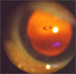
FALLS CHURCH, Va. — Some people call them floaters. Eye doctors call them “vitreous opacities.” Emily Flynn of Dunedin, New Zealand, called hers “a little fuzzball.” She flew halfway around the world to have it removed.

After more than 100 pinpoint zaps from a laser beam during a half-hour visit to a northern Virginia office park, the fuzzball was gone, obliterated within the clear, gelatinous goo that fills the eyeball.

The surgeon, John Karickhoff, has done the same procedure more than 1,400 times over the past 15 years and claims a success rate of better than 90%, with minimal risk of complications. Still, many ophthalmologists have never heard of the procedure — and most would recommend against it. The procedure has drawn regulatory scrutiny in Florida.

Nearly everybody has floaters or will develop them at some point in life, especially older and nearsighted people. Sometimes shaped like specks or snakes, they float through a person’s field of vision, and are most easily seen when you look against a light background like a blue sky or a white wall.

They are harmless, usually just bits of membrane that have become dislodged from other parts of the eye. Karickhoff estimated that 95% of people who have floaters ought to leave them alone. But for that 5%, they can be a legitimate problem.

“I’ve had musicians who couldn’t read the sheet music quickly enough because the floater would get in the way,” said Karickhoff, who has tried for years to get the procedure accepted into the medical mainstream. “It can ruin a patient’s quality of life.”

Only a handful of doctors in the United States — perhaps as few as two — regularly treat floaters with laser surgery.

“Patients usually are told to learn to live with it,” Karickhoff said. “By the time they come to see me, they’ve already seen two or three other doctors.”

Richard Bensinger, a spokesman for the American Academy of Ophthalmology, said most ophthalmologists believe the procedure is unnecessary. He acknowledged the laser treatment is often successful and carries little risk, but most doctors believe any risk is unacceptable for treating a benign condition like floaters.

If patients insist on treatment, Bensinger said the laser treatment can be a better choice than the more common alternative, a vitrectomy, which involves removing most or all of the eyeball’s internal fluid.

The vast majority of patients can learn to live with floaters, Bensinger said. Those patients who demand treatment, he said, “are mostly obsessive-compulsive types” who allow the floaters to drive them to distraction.

But Oliver Hill, 60, of Waynesburg, Pa., said many eye doctors are too dismissive of how irritating a floater can be. He compared it to trying to read a book while holding a pencil directly in front of one of your eyes.

“It just drove me right up the wall,” said Hill, who had his left eye treated in June 2005 and returned to Karickhoff in November to treat a floater in his right eye.

In Flynn’s case, her optometrist in New Zealand had told her that floaters were common and not harmful, and that she should learn to live with it. She learned about Karickhoff from his website.

Before having the surgery, Flynn said she would get headaches as she read and often tried to read with one eye shut to keep the floater out of her line of sight.

“That eye was effectively dysfunctional,” Flynn said.

Karickhoff said he knows firsthand that floaters can be troubling — he had what he called “a whopper” of a floater in his own eye.

He was skeptical when, on a Florida vacation, he saw a newspaper ad touting laser surgery for floaters. But he sat in on a surgery performed by Fort Myers ophthalmologist Scott Geller and later allowed Geller to operate on his eye. The procedure was a success.

Not long after, Karickhoff began performing the procedure himself. Both Geller and Karickhoff have since operated on thousands of patients.

Last year, the Florida Board of Medicine investigated a complaint against Geller filed by a 72-year-old woman who complained that the surgery merely broke up one large floater into a bunch of smaller floaters. But the board dismissed the complaint after hearing testimony from Karickhoff on how the procedure works.

Karickhoff, who has a clean record with the Virginia medical board, said he can tell by examining the patient’s eye if the floater is a serious distraction or a minor annoyance. He declines to treat those “who are making a mountain out of a mole hill” because such patients will almost never be satisfied.

There is little peer-reviewed literature about the procedure. What does exist shows the procedure to be generally safe, but the results about its effectiveness are mixed.

Karickhoff said studies have not replicated his success rate because they have not fully emulated his technique, which requires a laser beam of a specific intensity, and because they have not always chosen ideal candidates for surgery.

Karickhoff said there is a theoretical risk of serious complications, including a detached retina, but that he has never seen it happen. He said his own rate of complications is about a tenth of a percent, usually involving increased pressure within the eye.

Geller likened laser treatment of floaters to “an orphaned surgical procedure” that has been unable to overcome entrenched skepticism.

Despite that skepticism, Karickhoff said the medical community has not been completely unresponsive. He has written a medical book about the procedure, and said he received mostly positive feedback when he presented his work in November at the national conference of the American Academy of Ophthalmology. And insurers frequently cover the procedure, he said.

Manfred von Fricken, chief of ophthalmology at Inova Fairfax Hospital,said he has seen many of Karickhoff’s patients who have benefited from the surgery.

“It’s an innovative approach,” said von Fricken, who agrees that eye doctors are too often dismissive of patient complaints about floaters. “He’s thinking out of the box, and I think it’s pretty imaginative.”

Copyright 2007 The Associated Press. All rights reserved. This material may not be published, broadcast, rewritten or redistributed.

http://usatoday30.usatoday.com/news/health/2007-01-27-floaters_x.htm?csp=34

Eye Health and Retinal Detachment

Retinal detachment is a very serious eye condition that happens when the retina separates from the tissue around it. Since the retina can’t work properly under these conditions, you could permanently lose vision if the detached retina isn’t repaired promptly.

Who’s at Risk for a Detached Retina?

You’re more likely to get a detached retina if you:

  • Are severely nearsighted
  • Have had an eye injury or cataract surgery
  • Have a family history of retinal detachment

You may have heard the words “retinal tear,” too. That’s not the same as a retinal detachment.

  • Retinal tears often happen first. If fluid from within the eye passes through a retinal tear, that can separate the retina from its underlying tissue — and that’s retinal detachment. Retinal detachment may also happen with no warning. That’s more likely in elderly or very nearsighted people.

If you are unsure about your risk of retinal detachment, talk to your eye doctor.

What Are the Symptoms of a Detached Retina?

A detached retina doesn’t hurt, so look for these symptoms:

  • Flashes of light
  • Seeing “floaters” (small flecks or threads)
  • Darkening of your peripheral (side) vision

If you notice any of those symptoms, contact your eye doctor immediately.

How Is a Detached Retina Diagnosed?

Your health care provider would give you an eye exam, which would include dilating your eyes. That lets them see if your retina is detached.

Early diagnosis is key to preventing vision loss from a detached retina.

How Is a Detached Retina Treated?

There are many ways to treat a detached retina. These include:

  • Laser (thermal) or freezing (cryopexy). Both of these approaches can repair a tear in the retina if it is diagnosed early enough. This procedure is often done in the doctor’s office.
  • Pneumatic retinopexy. This procedure can be used to treat retinal detachment if the tear is small and easy to close. A small gas bubble is injected into the eye (specifically, into the  the clear, gel-like substance between the lens and the retina), where it then rises and presses against the retina, closing the tear. A laser or cryopexy can then be used to seal the tear.
  • Scleral buckle. This treatment for retinal detachment involves surgically sewing a silicone band (buckle) around the white of the eye (called the sclera) to push the sclera toward the tear until the tear heals. This band is not visible and remains permanently attached. Thermal treatment may then be necessary to seal the tear.
  • Vitrectomy. This surgery for retinal detachment is used for large tears. During a vitrectomy, the doctor removes the vitreous (the clear, gel-like substance between eye’s lens and retina) and replaces it with a saline solution.

Can a Detached Retina Be Prevented?

Yes, in some cases.

Getting an eye exam can flag early changes in your eyes that you may not have noticed. Treating those changes can help.

You should get your eyes checked once a year, or more often if you have conditions such as diabetes that make you more likely to have eye disease. Regular eye exams are also important if you are very nearsighted, as nearsightedness makes retinal detachment more likely.

If you have diabetes or high blood pressure, keeping those conditions under control will help the blood vessels in your retina, which is good for your eyes.

Not sure how often you should get your eyes checked? Ask your eye doctor.

You should also use the appropriate eye protection for certain activities. For instance, you should wear sports goggles with polycarbonate lenses while playing racquetball or certain other sports. You may also need eye protection if you work with machines, chemicals, or tools at work or home.

http://www.webmd.com/eye-health/eye-health-retinal-detachment

Computer Use and Eye Strain

Staring at your computer screen, smartphone, video game or other digital devices for long periods won’t cause permanent eye damage, but your eyes may feel dry and tired. Some people also experience headaches or motion sickness when viewing 3-D, which may indicate that the viewer has a problem with focusing or depth perception.

What causes computer-use eyestrain?

Woman looking at laptop
  • Normally, humans blink about 18 times a minute, but studies show we blink half that often while using computers and other digital screen devices, whether for work or play.
  • Extended reading, writing or other intensive “near work” can also cause eyestrain.

What to do:

  • Sit about 25 inches from the computer screen and position the screen so your eye gaze is slightly downward.
  • Reduce glare from the screen by lighting the area properly; use a screen filter if needed.
  • Post a note that says “Blink!” on the computer as a reminder.
  • Every 20 minutes, shift your eyes to look at an object at least 20 feet away, for at least 20 seconds: the “20-20-20” rule.
  • Use artificial tears to refresh your eyes when they feel dry.
  • Take regular breaks from computer work, and try to get enough sleep at night.

Computer-use eyestrain can be made worse by:

Sleep deprivation. When you get less sleep than you need, your eyes may become irritated. During sleep our eyes rest for an extended period and are replenished by nutrients. Ongoing eye irritation can lead to swelling and infection, especially if you wear contact lenses.

  • If you have to be at your computer for a marathon work session, take regular rest breaks or “power naps,” if possible.
  • Apply a washcloth soaked in warm water to tired, dry eyes (with eyes closed).
  • Use tired or sore eyes as a signal that it’s time to stop working and get some rest or sleep.

Incorrect contact lens use. If you wear contact lenses, it’s important that you use and care for them properly — especially if you use a computer and other digital-screen devices often. This helps avoid eye irritation, swelling, infection and vision problems.

  • Give your eyes a break: wear your glasses!
  • Don’t sleep in your contact lenses, even if they are labeled “extended wear.”
  • Always use good cleaning practices.

There are some important things to keep in mind when cleaning your contact lenses. You should:

  • Avoid touching the lenses with water; use fresh solution every time for cleaning and storing.
  • Rub your contacts when you clean them, even if you use a no-rub solution.
  • Clean your storage case regularly (with fresh solution, not water) and replace it every 2 to 3 months.

Stop wearing your contact lenses and see an ophthalmologist (Eye M.D.) right away if you develop any of these problems: Eyes that are red, blurry, watery, sensitive to light, or sore; eye swelling or discharge.

http://www.geteyesmart.org/eyesmart/living/computer-usage.cfm

Myths and Facts About Ophthalmology

Myth

Reading in dim light is harmful to your eyes.

Fact

Although reading in dim light can make your eyes feel tired, it is not harmful.

Myth

It is not harmful to watch a welder or look at the sun if you squint, or look through narrowed eyelids.

Fact

Even if you squint, ultra-violet light still gets to your eyes, damaging the cornea, lens, and retina. Never watch welding without wearing the proper protection. Never look directly at an eclipse.

Myth

Using a computer, or video display terminal (VDT), is harmful to the eyes.

Fact

Although using a VDT is associated with eyestrain or fatigue, it is not harmful to the eyes.

Myth

If you use your eyes too much, you wear them out.

Fact

You can use your eyes as much as you wish – they do not wear out.

Myth

Wearing poorly-fit glasses damages your eyes.

Fact

Although a good fit is required for good vision with glasses, a poor fit does not damage your eyes.

Myth

Wearing poorly fit contacts does not harm your eyes.

Fact

Poorly fit contact lenses can be harmful to your cornea (the window at the front of your eye). Make certain your eyes are checked regularly by an eye doctor at our ophthalmology office if you wear contact lenses.

Myth

You do not need to have your eyes checked until you are in your 40s or 50s.

Fact

There are several asymptomatic, yet treatable, eye diseases (most notably glaucoma) that can begin prior to your 40s.

Myth

Safety goggles are more trouble than they’re worth.

Fact

Safety goggles prevent many potentially blinding injuries every year. Keep goggles handy and use them.

Myth

It’s okay to swim while wearing soft contact lenses.

Fact

Potentially blinding eye infections can result from swimming or using a hot tub while wearing contact lenses.

Myth

Children outgrow crossed eyes.

Fact

Children do not outgrow truly crossed eyes. A child whose eyes are misaligned has strabismus and can develop poor vision in one eye (a condition known as amblyopia) because the brain turns off the misaligned or “lazy” eye. The sooner crossed or misaligned eyes are treated, the less likely the child will have permanently impaired vision.

Myth

A cataract must be ripe before it can be removed.

Fact

With modern cataract surgery, a cataract does not have to ripen before it is removed. When a cataract keeps you from doing the things you like or need to do, consider having it removed.

Myth

Cataracts can be removed with lasers.

Fact

A cataract cannot be removed with a laser. The cloudy lens must be removed through a surgical incision. However, after cataract surgery, a membrane within the eye may become cloudy. This membrane can be opened with laser eye surgery.

Myth

Eyes can be transplanted.

Fact

The eye cannot be transplanted. It is connected to the brain by the optic nerve, which cannot be reconnected once it has been severed. A person can, however, undergo cornea transplant surgery to replace the cornea, the clear part of the eye. Surgeons often use plastic intraocular lens implants (IOLs) such as crystalens®, ReSTOR®, ReZoom™ or TECNIS® to replace natural lenses during cataract surgery.

http://www.batravision.com/html/eye-care.html

Link


glasses-jpgBy Pure Matters

Imagine what life would be like if you couldn’t see well. Reading might be out. Watching a movie could be tough. Focusing on the face of a loved one could drive you to tears.

The number of people losing their vision is growing, yet experts say much of this vision loss could be prevented.

“We can intervene best when we identify a problem in the early stages,” says Roy S. Rubinfeld, M.D., a clinical correspondent for the American Academy of Ophthalmology (AAO). He warns against putting off regular eye exams because your eyes feel fine or you don’t wear glasses or contact lenses. Signs of some eye diseases, such as glaucoma and age-related macular degeneration (AMD), are present before you might notice symptoms.


Growing number


The National Eye Institute says more than 3.3 million Americans ages 40 and older have blindness or low vision. The institute projects that figure will increase markedly by the year 2020. The percentage of people more than 60 years old who suffer vision loss is growing fast, too.


“At 60, everyone should have an annual eye exam, even if you’re seeing very well,” Dr. Rubinfeld says.


Many diseases cause vision loss as we age, but AMD is the Western world’s top cause of blindness. Leading to loss of your central vision, it may cause dark spots in your sight, make straight lines appear wavy, or cause text to seem blurry. AMD, glaucoma, diabetic retinopathy, cataracts, and dry eye syndrome can all rob you of sight.


It’s best to see your eye doctor before trouble starts. But these signs should prompt a visit at once:


  • Trouble seeing objects close up or far away
  • Colors that seem faded
  • Poor night vision
  • Double or multiple vision
  • Loss of side vision
  • Poor central vision or straight objects that look wavy
  • Blurry text or type


Save aging eyes


Dr. Rubinfeld offers these recommendations:


Renee C. Bovelle, MD, FAAO, Represents Maryland with Entry into Renowned Publication The Leading Physicians of the World

Image

The Leading Physicians of the World and the International Association of Healthcare Professionals are proud to announce that Renee C. Bovelle, MD, FAAO, is being honored for her exceptional successes in the ophthalmology community.  Over 15 years practicing, Dr. Bovelle has already demonstrated the passion, dedication, and enthusiasm for patient care necessary to be considered a Top Physician in her field.

Dr. Bovelle currently maintains her own practice, Envision Eye and Laser Center, located in Glenn Dale, Maryland. Within her practice, Dr. Bovelle has performed thousands of LASIK, laser, ophthalmic, cataract and BOTOX procedures; with specific specialization in cornea and refractive surgery. Certified by the American Board of Ophthalmology, she offers both non-surgical and surgical treatments to enhance and rejuvenate her patients; as well as individualized treatment plans to help her patients make informed decisions about their medical, surgical, and aesthetic treatments. Her passion for the field goes beyond her practice, Dr. Bovelle’s work includes clinical research on modern devices used in modern refractive surgery; having her findings published in medical textbooks and ophthalmology journals in the United States and internationally. She also lectures across the globe. Dr. Bovelle also donates her time to the underprivileged both locally and internationally by participating in health fairs and medical mission trips including in Prince George’s County, Washington DC, Jamaica, Africa and India. Also being an advocate for continued education, Dr. Bovelle attends additional training courses in the disciplines of oculo-plastics and aesthetics; as well as, served as an Assistant Professor of Ophthalmology at Louisiana State University, and has worked at the National Institutes of Health.

Dr. Bovelle’s career began in 1992, after she received her medical degree from the University of California in Los Angeles. She completed her internships at a Johns Hopkins-affiliated hospital and at the MedStar Franklin Square Medical Center.  Her residency in ophthalmology was completed at the esteemed Yale University, where she served as chief resident. Upon completion, Dr. Bovelle attended the Louisiana State University Eye Center for a Cornea and Refractive Surgery fellowship under the direction of Dr. Herbert Kaufman, one of the world’s leading corneal and refractive surgeons.

To find out more about Dr. Bovelle, please visit www.renee-bovelle.findatopdoc.com andwww.envisioneyeandlaser.com.  Be sure to look out for her upcoming publication in The Leading Physicians of the World.