Monthly Archives: August 2014

DNA test for congenital cataracts leads to faster, more accurate diagnoses of rare diseases linked to childhood blindness

Ophthalmology_Pediatrics

Researchers in the United Kingdom have demonstrated that advanced DNA testing for congenital cataracts can quickly and accurately diagnose a number of rare diseases marked by childhood blindness, according to a study published online in Ophthalmology, the journal of the American Academy of Ophthalmology. Using a single test, doctors were able to tailor care specifically to a child’s condition based on their mutations reducing the time and money spent on diagnosis and enabling earlier treatment and genetic counseling.

Each year, between 20,000 and 40,000 children worldwide are born with congenital cataracts, a disease that clouds the lens of the eye and often requires surgery and treatment to prevent blindness.[1] The disease can arise following a maternal infection or be inherited as an isolated abnormality. Congenital cataracts can also appear as a symptom of more than 100 rare diseases, making mutations in the 115 genes associated with congenital cataracts useful as diagnostic markers for the illnesses.

Diagnosing these rare diseases previously proved a lengthy, costly and inconclusive process involving numerous clinical assessments and taking a detailed family history. DNA testing, one gene at a time, would have taken years to complete. Employing new DNA sequencing technology, called targeted next-generation sequencing, researchers at the University of Manchester sped up diagnosis to a matter of weeks by testing for mutations in all 115 known congenital cataracts genes at one time.

In 75 percent of the 36 cases tested, the DNA test determined the exact genetic cause of congenital cataracts. In one case, the DNA test helped diagnose a patient with Warburg Micro syndrome, an extremely rare disease that is marked by an abnormally small head and the development of severe epilepsy, among other medical issues. Having a clear diagnosis allowed for genetic counseling and appropriate care to be delivered quicker than previously possible without the test.

“There are many diseases that involve congenital cataracts but finding the exact reason was always difficult,” said Graeme Black, DPhil., professor of genetics and ophthalmology at the University of Manchester and strategic director of the Manchester Centre for Genomic Medicine. “Even with a family history, diagnosing these rare diseases was always a bit of a shot in the dark.”

In the course of their work, done in collaboration with Manchester Royal Eye Hospital, researchers also found previously undescribed mutations linked to cataract formation. “There is hope that our work may one day provide more insight into the development and treatment of age-related cataracts, a leading cause of blindness worldwide,” said Rachel Gillespie, MSc, lead author of the study who designed and developed the test.

The test was made available to U.K. patients through the country’s National Health Service in December 2013. Infants and children who have congenital cataracts can be tested as well as prospective parents with a history of the condition who wish to evaluate the risk to their child. Results generally take about two months. While only available in the U.K., the congenital cataract DNA test can be requested by registered medical facilities through international referral.

As with all genetic testing, the American Academy of Ophthalmology encourages clinicians and patients to consider the benefits as well as the risks. Ophthalmologists who order genetic tests either should provide genetic counseling to their patients themselves, if qualified to do so, or should ensure that counseling is provided by a trained individual, such as a board-certified medical geneticist or genetic counselor. For more information, please see the Academy’s recommendations on genetic testing for inherited eye diseases.


http://www.medicalnewstoday.com/releases/281442.php


Slippery material for lubricating joints inspired by nature

Ophthalmology_Orthopedic Surgery

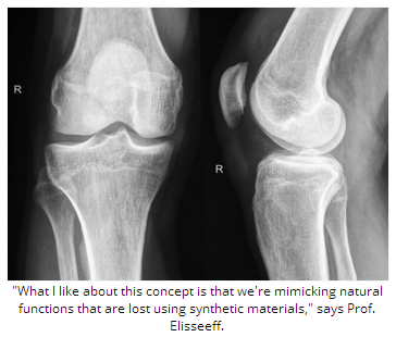
For some time, scientists have been aware that synovial fluid in joints contains a natural substance that helps keep them well lubricated. Now, by mimicking its properties, engineers at Johns Hopkins University in Baltimore, MD, hope to develop a new material that delivers long-lasting lubrication in artificial joints, and other specific spots in the body where surfaces move against each other.

The team writes about the new material, and the potential it offers, in the journal Nature Materials. As well as helping to ease joint pain, other areas of possible use include making contact lenses more comfortable.

The molecule they are investigating is called hyaluronic acid (HA), which exists in several forms in the body where lubrication is needed.

One form of HA reduces inflammation and protects cells from metabolic damage. In the body, HA is bound to the surfaces it protects by a protein. Research shows that in damaged, diseased and aging knees, hips, shoulders and elbows, this protein is no longer able to hold onto HA.

Viscosupplementation is a popular treatment for painful joints and consists of injecting HA into the painful joint. However, if the cause of the pain is the lack of the protein that helps bind HA to the affected surface, then it does not offer long-lasting benefit. The injected HA is soon washed away by the body’s natural cleaning processes.

‘Chemical handle’ holds HA in place

Thus, led by Jennifer H. Elisseeff, a professor at the Wilmer Eye Institute at Johns Hopkins, the team sought to find a way to keep HA in place. They found their answer in molecules known as HA-binding peptides (HABpeps).

The scientists used HABpep as a “chemical handle” to attach HA onto natural and artificial surfaces with the help of another synthetic molecule, polyethylene glycol.

In the lab, they tested the new material in cultured tissue and joint and eye surface tissue in live animals. They found the bound HA did not wash away easily, and it reduced friction as well as when the tissues were immersed in a bath of HA:

“Tissue surfaces treated with the HA-binding system exhibited higher lubricity values, and in vivo were able to retain HA in the articular joint and to bind ocular tissue surfaces,” they note.

HABpep holds HA in place 12 times longer

In another set of experiments, they also tested an HABpep designed to attach to cartilage. They injected rats’ knees first with some HABpep, then with some HA, and found the HA stayed in place 12 times longer than it did in rats that had received only HA injections without HABpep.

The team suggests this shows HABpeps may be a useful addition to viscosupplementation by helping the HA to stay in place longer. They also conclude:

“Biomaterials-mediated strategies that locally bind and concentrate HA could provide physical and biological benefits when used to treat tissue-lubricating dysfunction and to coat medical devices.”

Although it will be some time before such a material is ready for use in humans, the team highlights that theirs is yet another example of where nature has inspired the solution to a medical problem.

Prof. Elisseeff, who is also of the Johns Hopkins University departments of Biomedical Engineering and of Materials Science and Engineering, says:

“What I like about this concept is that we’re mimicking natural functions that are lost using synthetic materials.”

Meanwhile in November 2013, Medical News Today learned how another team of researchers – also inspired by nature – is refining sea coral for use in bone grafts.

Written by Catharine Paddock PhD

http://www.medicalnewstoday.com/articles/281212.php