Monthly Archives: July 2013

Corneal Abrasions

ImageWhat is a corneal abrasion?

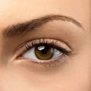
A corneal abrasion is a cut or scratch on your cornea. The cornea is a clear layer of protective tissue at the front of your eye that lies over the iris (which is the colored part of your eye). The cornea helps focus light.

What can cause a corneal abrasion?

A corneal abrasion can occur when something gets into your eye, such as sand, dust, dirt, and wood or metal shavings. The cornea can also be scratched by a fingernail, a tree branch or a contact lens that is dry or dirty. Rubbing your eyes very hard is another way that an abrasion can occur.

In some people, the outer layers of the cornea are weak. These people may get a corneal abrasion for no apparent reason.

How do I know if I have a corneal abrasion?

The cornea is very sensitive, and a corneal abrasion is usually quite painful. You may feel like you have sand or grit in your eye. You may notice tears or blurred vision, or your eye may look red. You may also notice that light hurts your eye. Some people get a headache when they have a corneal abrasion.

What do I do if I get something in my eye?

If you think something has gotten into your eye, first try to wash out the eye by flushing it with clean water or saline solution. Your workplace may have an eye rinse station for this purpose. Sometimes, blinking or pulling the upper eyelid over the lower eyelid may remove a particle from under the eyelid. Avoid rubbing your eye. If you or someone else notices something on the white part of your eye, use a soft tissue or cotton swab to gently lift it out of the eye. Don’t try to remove something that is directly over the cornea — this might cause more serious damage. Call your doctor if you can’t remove the particle or if there doesn’t seem to be anything in your eye.

What will my doctor do for a corneal abrasion?

Your doctor will examine your eye for any damage or particles that may be trapped under your eyelid. A yellow-orange dye may be placed on your eye to help your doctor see the abrasion. Your doctor will probably treat the abrasion with eye drops or ointment. Most small abrasions heal within one to three days. You may need to return to your doctor for another exam the next day.

What if I wear contact lenses?

If you wear contact lenses, you need to be especially careful with a corneal abrasion because you have a higher risk of infection. Your doctor may tell you not to wear your contact lenses for a few days after a corneal abrasion, especially if you’re treating your eye with medicated drops.

How can I prevent corneal abrasion?

Take the following steps to help prevent corneal abrasions:

  • Wear protective eye goggles when you’re around machinery that causes particles of wood, metal or other materials to fly into the air (such as a chainsaw or a sandblaster).
  • Keep your fingernails trimmed short. Cut infants’ and young children’s fingernails short, also.
  • Trim low-hanging tree branches.
  • Use care when putting in contact lenses, and make sure you clean them properly each day.
  • Don’t sleep in your contact lenses.

http://familydoctor.org/familydoctor/en/prevention-wellness/staying-healthy/first-aid/corneal-abrasions.html

What is an Ophthalmologist?


When you go to “get your eyes checked,” there are a variety of eye care providers you might see. Ophthalmologists, optometrists and opticians all play an important role in providing eye care services to consumers. However, each group has different levels of training and expertise; you should be sure you are seeing the right provider for your condition or treatment.

What is an ophthalmologist?

An ophthalmologist – Eye M.D. – is a medical or osteopathic doctor who specializes in eye and vision care. Ophthalmologists are specially trained to provide the full spectrum of eye care, from prescribing glasses and contact lenses to complex and delicate eye surgery. Many ophthalmologists are also involved in scientific research into the causes and cures for eye diseases and vision problems.

How is an ophthalmologist different from an optometrist and an optician?

Ophthalmologists are different from optometrists and opticians in their training and in what they can diagnose and treat. As a medical doctor, an ophthalmologist is licensed to practice medicine and surgery. An ophthalmologist diagnoses and treats all eye diseases, performs eye surgery and prescribes and fits eyeglasses and contact lenses. Ophthalmologists complete:

•  four years of college;

•  four years of medical school;

•  one year of internship;

•  three years, at least, of residency (hospital-based training) in the diagnosis and medical and surgical treatment of eye disorders.

An optometrist receives a Doctor of Optometry (OD) degree and is licensed to practice optometry, not medicine. The practice of optometry traditionally involves examining the eye for the purpose of prescribing and dispensing corrective lenses, screening vision to detect certain eye abnormalities, and prescribing medications for certain eye diseases.

An optician is trained to design, verify and fit eyeglass lenses and frames, contact lenses, and other devices to correct eyesight. They use prescriptions supplied by ophthalmologists or optometrists, but do not test vision or write prescriptions for visual corrections. Opticians are not permitted to diagnose or treat eye diseases.

How does an ophthalmologist become certified?

After four years of college and eight additional years of medical education and training, a certified ophthalmologist must pass a rigorous two-part examination given by the American Board of Ophthalmology.

http://www.eyecareamerica.org/eyecare/tmp/what-is-an-ophthalmologist.cfm

Scientist Working To Break Vicious Cycle Causing Vision Loss In Diabetes


It’s a vicious cycle that robs people with diabetes of their vision.

The hallmark high glucose of the disease causes inflammation that produces free radicals that cause inflammation that produces more free radicals, explains Dr. Manuela Bartoli, vision scientist at the Medical College of Georgia at Georgia Regents University.

If that’s not bad enough, the body’s endogenous system for dealing with free radicals also is dramatically impacted by diabetes, said Bartoli, who recently received a $1.8 million grant from the National Eye Institute to try to bolster that system and interrupt the destructive cycle.

Nearly 10 percent of the U.S. population has diabetes, according to the National Diabetes Foundation, and nearly half those individuals will develop diabetic retinopathy, according to the National Eye Institute.

Culprit free radicals are actually normal byproducts of the body’s constant use of oxygen and, despite their derivative status, also are important signaling molecules in the body. Problems result when there are too many, like in diabetes, and their natural tendency to bond starts wreaking havoc on cells and DNA. In fact, excessive levels are thought to be a major contributor to a wide variety of diseases as well as aging.

The thioredoxin system typically works to maintain a healthy level of free radicals by neutralizing excess but, like many body systems, the thioredoxin system slows with age and diabetes hastens the process.

“This increase in free radicals results in an inability to put them to good use,” Bartoli said. “Instead, we accumulate the damage they induce.” In the case of the eyes and diabetes, over time the overwhelmed system destroys blood vessels that deliver blood and nutrition. In another biological irony, the starving eyes grow new blood vessels but they are fragile, leaky and often misplaced so ultimately they destroy vision.

Bartoli believes a selenium supplement could give the thioredoxin system the shot in the arm needed to stay efficient and effective. Selenium is a byproduct itself, resulting from copper-refining and used to make glass, alloys and more. It is also found in fish, nuts and grains.

Thioredoxin reductase, a protein essential to the recycling of the system, is dependent on selenium and Bartoli has found that protein’s activity is reduced in an animal model of diabetic retinopathy and in retinas of human diabetic donors. Bartoli believes the cascade of cellular change resulting from high glucose levels impairs thioredoxin reductase. So she wants to better understand how the system works, exactly what happens to thioredoxin reductase and whether supplements of selenium can help the natural antioxidant system work better in diabetes.

In a related study, funded by the International Retinal Research Foundation, she is looking for an early sign of eye damage and possibly another window of intervention.

Currently, swelling of the macula – the central part of the retina responsible for central vision – is the first sign of treatable trouble. Anti-inflammatories injected into the eyes can help.

However increased blood levels of uric acid, a part of the inflammatory process that leads to swelling, may be an earlier indicator, Bartoli said. Uric acid is a byproduct of purine metabolism and is typically eliminated in the urine. High uric acid levels are associated with cardiovascular disease and gout as well as diabetes but it hasn’t been well studied in the eye.

“We want to validate hyperuricemia as a risk factor for progression of diabetic retinopathy,” she said. So she and her colleagues are measuring levels in the blood and eye fluid to see if they correlate with each other and with progressive eye damage. They also are reducing uric acid levels by giving two drugs already on the market, one that blocks formation and another that enhances excretion. Thinking that uric acid levels also may be a biomarker, she eventually wants to see how uric acid levels correlate with disease progression in humans.

“As the ancients said: ‘The eyes are the mirror of the soul.’ We also know that whatever happens in the eye is an expression of what is happening in the rest of the body,” Bartoli said. “We want to better understand the causes of inflammation in the eye in diabetes and find better ways to manage it as well as byproducts such as uric acid. Ultimately, of course, we hope to protect sight.”

 
http://www.medicalnewstoday.com/releases/262731.php